 Movie
Movie Controller

[English] 日本語
 Yorodumi
- PDB-5xmo: X-ray crystal structure of Pseudoazurin Met16Phe/Thr36Lys variant -
+ Open data

- Basic information
| Entry | Database: PDB / ID: 5xmo | ||||||
|---|---|---|---|---|---|---|---|
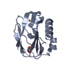
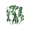
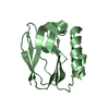
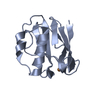
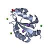
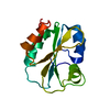
| Title | X-ray crystal structure of Pseudoazurin Met16Phe/Thr36Lys variant | ||||||
 Components Components | Pseudoazurin | ||||||
 Keywords Keywords |  ELECTRON TRANSPORT / Electron transfer / Cupredoxin ELECTRON TRANSPORT / Electron transfer / Cupredoxin | ||||||
| Function / homology |  Function and homology information Function and homology information | ||||||
| Biological species |  Achromobacter cycloclastes (bacteria) Achromobacter cycloclastes (bacteria) | ||||||
| Method | X-RAY DIFFRACTION / SYNCHROTRON / MOLECULAR REPLACEMENT / molecular replacement / Resolution: 1.19 Å | ||||||
 Authors Authors | Yamaguchi, T. / Kohzuma, T. | ||||||
| Funding support |  Japan, 1items Japan, 1items
| ||||||
 Citation Citation | Journal: Photon Factory Activity Report / Year: 2017 Title: X-ray Crystallographic Analysis of M16F/T36K Double Mutant of Pseudoazurin Authors: Yamaguchi, T. / Takebayashi, N. / Akao, K. / Sakai, C. / Kohzuma, T. | ||||||
| History |
|
- Structure visualization
| Structure viewer | Molecule: MolmilJmol/JSmol |
|---|
- Downloads & links
-Download
| PDBx/mmCIF format | 5xmo.cif.gz | 75.7 KB | Display | PDBx/mmCIF format |
|---|---|---|---|---|
| PDB format | pdb5xmo.ent.gz | 55.2 KB | Display | PDB format |
| PDBx/mmJSON format | 5xmo.json.gz | Tree view | PDBx/mmJSON format | |
| Others |  Other downloads Other downloads |
-Validation report
| Arichive directory | https://data.pdbj.org/pub/pdb/validation_reports/xm/5xmoftp://data.pdbj.org/pub/pdb/validation_reports/xm/5xmo | HTTPS FTP |
|---|
-Related structure data
| Related structure data |  1bqkS S: Starting model for refinement |
|---|---|
| Similar structure data |


-Links
 PDBj
PDBj- Assembly
| Deposited unit | 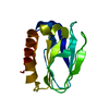
| ||||||||
|---|---|---|---|---|---|---|---|---|---|
| 1 |
| ||||||||
| Unit cell |
| ||||||||
| Components on special symmetry positions |
|
-Components
| #1: Protein | Mass: 13076.994 Da / Num. of mol.: 1 / Mutation: M16F, T36K Source method: isolated from a genetically manipulated source Source: (gene. exp.) Achromobacter cycloclastes (bacteria) / Gene: bcp / Production host: Escherichia coli (E. coli) / References: UniProt: P19567 |
|---|---|
| #2: Chemical | ChemComp-CU / Copper  Mass: 63.546 Da / Num. of mol.: 1 / Source method: obtained synthetically / Formula: Cu Mass: 63.546 Da / Num. of mol.: 1 / Source method: obtained synthetically / Formula: Cu |
| #3: Water | ChemComp-HOH / Water Mass: 18.015 Da / Num. of mol.: 208 / Source method: isolated from a natural source / Formula: H2O Mass: 18.015 Da / Num. of mol.: 208 / Source method: isolated from a natural source / Formula: H2O |
-Experimental details
-Experiment
| Experiment | Method: X-RAY DIFFRACTION / Number of used crystals: 1 |
|---|
- Sample preparation
| Crystal | Density Matthews: 2.52 Å3/Da / Density % sol: 51.27 % |
|---|---|
| Crystal grow | Temperature: 293 K / Method: vapor diffusion, sitting drop / pH: 7.5 Details: Buffer for crystallization: 0.1 M sodium acetate pH 4.5 Precipitant: 30 % PEG1500 Soaking (Cryoprotectant): 0.1 M Tris-HCl pH 7.5, 40 % PEG1500 |
-Data collection
| Diffraction | Mean temperature: 100 K |
|---|---|
| Diffraction source | Source: SYNCHROTRON / Site: Photon Factory / Beamline: AR-NW12A / Wavelength: 1 Å |
| Detector | Type: ADSC QUANTUM 210 / Detector: CCD / Date: May 6, 2017 |
| Radiation | Protocol: SINGLE WAVELENGTH / Monochromatic (M) / Laue (L): M / Scattering type: x-ray |
| Radiation wavelength | Wavelength: 1 Å / Relative weight: 1 |
| Reflection | Resolution: 1.192→46.048 Å / Num. obs: 38384 / % possible obs: 92.3 % / Redundancy: 5.4 % / Rpim(I) all: 0.033 / Rrim(I) all: 0.079 / Rsym value: 0.072 / Net I/av σ(I): 6.8 / Net I/σ(I): 12.5 |
| Reflection shell | Resolution: 1.192→1.223 Å / Redundancy: 5.4 % / Rmerge(I) obs: 0.317 / Mean I/σ(I) obs: 4.4 / % possible all: 87.3 |
-Phasing
| Phasing | Method: molecular replacement | ||||||
|---|---|---|---|---|---|---|---|
| Phasing MR | R rigid body: 0.517
|
- Processing
| Software |
| ||||||||||||||||||||||||||||||||||||||||||||||||||||||||||||||||||||||||||||||||||||||||||||||||||||||||||||||||||||||||||||||||||||||||||||||||||||||||||||||||||||||||||||||||||||||
|---|---|---|---|---|---|---|---|---|---|---|---|---|---|---|---|---|---|---|---|---|---|---|---|---|---|---|---|---|---|---|---|---|---|---|---|---|---|---|---|---|---|---|---|---|---|---|---|---|---|---|---|---|---|---|---|---|---|---|---|---|---|---|---|---|---|---|---|---|---|---|---|---|---|---|---|---|---|---|---|---|---|---|---|---|---|---|---|---|---|---|---|---|---|---|---|---|---|---|---|---|---|---|---|---|---|---|---|---|---|---|---|---|---|---|---|---|---|---|---|---|---|---|---|---|---|---|---|---|---|---|---|---|---|---|---|---|---|---|---|---|---|---|---|---|---|---|---|---|---|---|---|---|---|---|---|---|---|---|---|---|---|---|---|---|---|---|---|---|---|---|---|---|---|---|---|---|---|---|---|---|---|---|---|
| Refinement | Method to determine structure: MOLECULAR REPLACEMENT Starting model: 1BQK Resolution: 1.19→46.05 Å / Cor.coef. Fo:Fc: 0.973 / Cor.coef. Fo:Fc free: 0.964 / SU B: 1.157 / SU ML: 0.023 / Cross valid method: THROUGHOUT / ESU R: 0.035 / ESU R Free: 0.037 / Stereochemistry target values: MAXIMUM LIKELIHOOD / Details: HYDROGENS HAVE BEEN ADDED IN THE RIDING POSITIONS
| ||||||||||||||||||||||||||||||||||||||||||||||||||||||||||||||||||||||||||||||||||||||||||||||||||||||||||||||||||||||||||||||||||||||||||||||||||||||||||||||||||||||||||||||||||||||
| Solvent computation | Ion probe radii: 0.8 Å / Shrinkage radii: 0.8 Å / VDW probe radii: 1.2 Å / Solvent model: MASK | ||||||||||||||||||||||||||||||||||||||||||||||||||||||||||||||||||||||||||||||||||||||||||||||||||||||||||||||||||||||||||||||||||||||||||||||||||||||||||||||||||||||||||||||||||||||
| Displacement parameters | Biso mean: 13.494 Å2
| ||||||||||||||||||||||||||||||||||||||||||||||||||||||||||||||||||||||||||||||||||||||||||||||||||||||||||||||||||||||||||||||||||||||||||||||||||||||||||||||||||||||||||||||||||||||
| Refinement step | Cycle: 1 / Resolution: 1.19→46.05 Å
| ||||||||||||||||||||||||||||||||||||||||||||||||||||||||||||||||||||||||||||||||||||||||||||||||||||||||||||||||||||||||||||||||||||||||||||||||||||||||||||||||||||||||||||||||||||||
| Refine LS restraints |
|