 Movie
Movie Controller

[English] 日本語
 Yorodumi
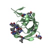
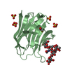
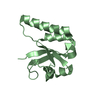
- PDB-5wwx: Crystal structure of the KH2 domain of human RNA-binding E3 ubiqu... -
+ Open data

- Basic information
| Entry | Database: PDB / ID: 5wwx | ||||||
|---|---|---|---|---|---|---|---|
| Title | Crystal structure of the KH2 domain of human RNA-binding E3 ubiquitin-protein ligase MEX-3C complex with RNA | ||||||
 Components Components |
| ||||||
 Keywords Keywords | RNA BINDING PROTEIN/RNA / KH2 / MEX-3C / RNA / RNA BINDING PROTEIN-RNA complex | ||||||
| Function / homology |  Function and homology information Function and homology informationchondrocyte hypertrophy / regulation of fat cell differentiation / energy homeostasis / RING-type E3 ubiquitin transferase / ubiquitin protein ligase activity / Antigen processing: Ubiquitination & Proteasome degradation / RNA binding / nucleus / metal ion binding / cytoplasm Similarity search - Function | ||||||
| Biological species |  Homo sapiens (human) Homo sapiens (human)synthetic construct (others) | ||||||
| Method |  X-RAY DIFFRACTION / SYNCHROTRON / MOLECULAR REPLACEMENT / Resolution: 2 Å X-RAY DIFFRACTION / SYNCHROTRON / MOLECULAR REPLACEMENT / Resolution: 2 Å | ||||||
 Authors Authors | Yang, L. / Wang, C. / Li, F. / Gong, Q. | ||||||
 Citation Citation | Journal: J. Biol. Chem. / Year: 2017 Title: The human RNA-binding protein and E3 ligase MEX-3C binds the MEX-3-recognition element (MRE) motif with high affinity Authors: Yang, L. / Wang, C. / Li, F. / Zhang, J. / Nayab, A. / Wu, J. / Shi, Y. / Gong, Q. | ||||||
| History |
|
- Structure visualization
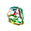
| Structure viewer | Molecule: MolmilJmol/JSmol |
|---|
- Downloads & links
-Download
| PDBx/mmCIF format | 5wwx.cif.gz | 37.3 KB | Display | PDBx/mmCIF format |
|---|---|---|---|---|
| PDB format | pdb5wwx.ent.gz | 22.9 KB | Display | PDB format |
| PDBx/mmJSON format | 5wwx.json.gz | Tree view | PDBx/mmJSON format | |
| Others |  Other downloads Other downloads |
-Validation report
| Summary document | 5wwx_validation.pdf.gz | 444.2 KB | Display | wwPDB validaton report |
|---|---|---|---|---|
| Full document | 5wwx_full_validation.pdf.gz | 444.2 KB | Display | |
| Data in XML | 5wwx_validation.xml.gz | 6.5 KB | Display | |
| Data in CIF | 5wwx_validation.cif.gz | 8.1 KB | Display | |
| Arichive directory | https://data.pdbj.org/pub/pdb/validation_reports/ww/5wwxftp://data.pdbj.org/pub/pdb/validation_reports/ww/5wwx | HTTPS FTP |
-Related structure data
| Related structure data |  5wwwSC  5wwzC S: Starting model for refinement C: citing same article ( |
|---|---|
| Similar structure data |


-Links
 PDBj
PDBj


- Assembly
| Deposited unit | 
| |||||||||
|---|---|---|---|---|---|---|---|---|---|---|
| 1 |
| |||||||||
| Unit cell |
| |||||||||
| Components on special symmetry positions |
|
-Components
| #1: Protein | Mass: 9772.222 Da / Num. of mol.: 1 / Fragment: KH2 domain, UNP residues 320-396 Source method: isolated from a genetically manipulated source Source: (gene. exp.) Homo sapiens (human) / Gene: MEX3C, RKHD2, RNF194, BM-013 / Production host:  References: UniProt: Q5U5Q3, RING-type E3 ubiquitin transferase |
|---|---|
| #2: RNA chain | Mass: 1610.032 Da / Num. of mol.: 1 / Source method: obtained synthetically / Source: (synth.) synthetic construct (others) |
| #3: Chemical | ChemComp-NI /   Mass: 58.693 Da / Num. of mol.: 1 / Source method: obtained synthetically / Formula: Ni Mass: 58.693 Da / Num. of mol.: 1 / Source method: obtained synthetically / Formula: Ni |
| #4: Water | ChemComp-HOH /  Mass: 18.015 Da / Num. of mol.: 62 / Source method: isolated from a natural source / Formula: H2O Mass: 18.015 Da / Num. of mol.: 62 / Source method: isolated from a natural source / Formula: H2O |
-Experimental details
-Experiment
| Experiment | Method: X-RAY DIFFRACTION / Number of used crystals: 1 |
|---|
- Sample preparation
| Crystal | Density Matthews: 2.02 Å3/Da / Density % sol: 39.18 % |
|---|---|
| Crystal grow | Temperature: 293 K / Method: vapor diffusion, sitting drop Details: 0.04M Magnesium acetate tetrahydrate, 0.05M Sodium cacodylate trihydrate pH 6.0, 30% v/v (+/-)-2-Methyl-2,4-pentanediol |
-Data collection
| Diffraction | Mean temperature: 100 K | ||||||||||||||||||||||||||||||||||||||||||||||||||||||||||||||||||
|---|---|---|---|---|---|---|---|---|---|---|---|---|---|---|---|---|---|---|---|---|---|---|---|---|---|---|---|---|---|---|---|---|---|---|---|---|---|---|---|---|---|---|---|---|---|---|---|---|---|---|---|---|---|---|---|---|---|---|---|---|---|---|---|---|---|---|---|
| Diffraction source | Source: SYNCHROTRON / Site: SSRF  / Beamline: BL18U1 / Wavelength: 0.979 Å / Beamline: BL18U1 / Wavelength: 0.979 Å | ||||||||||||||||||||||||||||||||||||||||||||||||||||||||||||||||||
| Detector | Type: ADSC QUANTUM 315r / Detector: CCD / Date: Dec 12, 2015 | ||||||||||||||||||||||||||||||||||||||||||||||||||||||||||||||||||
| Radiation | Protocol: SINGLE WAVELENGTH / Monochromatic (M) / Laue (L): M / Scattering type: x-ray | ||||||||||||||||||||||||||||||||||||||||||||||||||||||||||||||||||
| Radiation wavelength | Wavelength: 0.979 Å / Relative weight: 1 | ||||||||||||||||||||||||||||||||||||||||||||||||||||||||||||||||||
| Reflection | Resolution: 2→40 Å / Num. obs: 6609 / % possible obs: 99.6 % / Redundancy: 7.6 % / Biso Wilson estimate: 23.13 Å2 / Rmerge(I) obs: 0.108 / Net I/σ(I): 3.6 | ||||||||||||||||||||||||||||||||||||||||||||||||||||||||||||||||||
| Reflection shell |
|
- Processing
| Software |
| ||||||||||||||||||||||||
|---|---|---|---|---|---|---|---|---|---|---|---|---|---|---|---|---|---|---|---|---|---|---|---|---|---|
| Refinement | Method to determine structure: MOLECULAR REPLACEMENT Starting model: 5WWW Resolution: 2→33.367 Å / SU ML: 0.23 / Cross valid method: FREE R-VALUE / σ(F): 1.37 / Phase error: 18.85
| ||||||||||||||||||||||||
| Solvent computation | Shrinkage radii: 0.9 Å / VDW probe radii: 1.11 Å | ||||||||||||||||||||||||
| Displacement parameters | Biso max: 106.81 Å2 / Biso mean: 23.0942 Å2 / Biso min: 8.76 Å2 | ||||||||||||||||||||||||
| Refinement step | Cycle: final / Resolution: 2→33.367 Å
| ||||||||||||||||||||||||
| Refine LS restraints |
| ||||||||||||||||||||||||
| LS refinement shell | Refine-ID: X-RAY DIFFRACTION / Rfactor Rfree error: 0 / Total num. of bins used: 2
|
