 Movie
Movie Controller

[English] 日本語
 Yorodumi
- PDB-5jli: Nitric oxide complex of the L16A mutant of cytochrome c prime fro... -
+ Open data

- Basic information
| Entry | Database: PDB / ID: 5jli | |||||||||
|---|---|---|---|---|---|---|---|---|---|---|
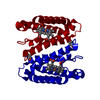
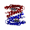
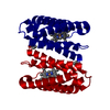
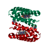
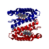
| Title | Nitric oxide complex of the L16A mutant of cytochrome c prime from Alcaligenes xylosoxidans | |||||||||
 Components Components | Cytochrome c' | |||||||||
 Keywords Keywords | ELECTRON TRANSPORT / gas sensor cytochrome nitric oxide | |||||||||
| Function / homology |  Function and homology information Function and homology informationelectron transport chain / electron transfer activity / periplasmic space / iron ion binding / heme binding Similarity search - Function | |||||||||
| Biological species |  Alcaligenes xylosoxydans xylosoxydans (bacteria) Alcaligenes xylosoxydans xylosoxydans (bacteria) | |||||||||
| Method |  X-RAY DIFFRACTION / SYNCHROTRON / MOLECULAR REPLACEMENT / Resolution: 1.55 Å X-RAY DIFFRACTION / SYNCHROTRON / MOLECULAR REPLACEMENT / Resolution: 1.55 Å | |||||||||
 Authors Authors | Kekilli, D. / Strange, R.W. / Hough, M.A. | |||||||||
 Citation Citation | Journal: Chem Sci / Year: 2017 Title: Engineering proximal vs. distal heme-NO coordination via dinitrosyl dynamics: implications for NO sensor design. Authors: Kekilli, D. / Petersen, C.A. / Pixton, D.A. / Ghafoor, D.D. / Abdullah, G.H. / Dworkowski, F.S.N. / Wilson, M.T. / Heyes, D.J. / Hardman, S.J.O. / Murphy, L.M. / Strange, R.W. / Scrutton, N. ...Authors: Kekilli, D. / Petersen, C.A. / Pixton, D.A. / Ghafoor, D.D. / Abdullah, G.H. / Dworkowski, F.S.N. / Wilson, M.T. / Heyes, D.J. / Hardman, S.J.O. / Murphy, L.M. / Strange, R.W. / Scrutton, N.S. / Andrew, C.R. / Hough, M.A. | |||||||||
| History |
|
- Structure visualization
| Structure viewer | Molecule: MolmilJmol/JSmol |
|---|
- Downloads & links
-Download
| PDBx/mmCIF format | 5jli.cif.gz | 76.6 KB | Display | PDBx/mmCIF format |
|---|---|---|---|---|
| PDB format | pdb5jli.ent.gz | 56.3 KB | Display | PDB format |
| PDBx/mmJSON format | 5jli.json.gz | Tree view | PDBx/mmJSON format | |
| Others |  Other downloads Other downloads |
-Validation report
| Arichive directory | https://data.pdbj.org/pub/pdb/validation_reports/jl/5jliftp://data.pdbj.org/pub/pdb/validation_reports/jl/5jli | HTTPS FTP |
|---|
-Related structure data
| Related structure data | 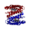 5jp7C 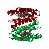 5jraC 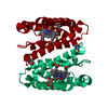 5js5C 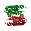 5jslC 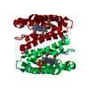 5jt4C  5juaC 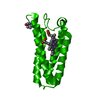 5jveC 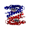 2yl0S S: Starting model for refinement C: citing same article ( |
|---|---|
| Similar structure data |


-Links
 PDBj
PDBj


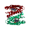
- Assembly
| Deposited unit | 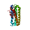
| ||||||||
|---|---|---|---|---|---|---|---|---|---|
| 1 | 
| ||||||||
| Unit cell |
| ||||||||
| Components on special symmetry positions |
|
-Components
-Protein / Sugars , 2 types, 2 molecules A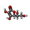
| #1: Protein | Mass: 13589.362 Da / Num. of mol.: 1 / Mutation: L16A Source method: isolated from a genetically manipulated source Source: (gene. exp.) Alcaligenes xylosoxydans xylosoxydans (bacteria)Plasmid: pET26b+ / Production host: |
|---|---|
| #3: Sugar | ChemComp-ASC /  Type: L-saccharide / Mass: 176.124 Da / Num. of mol.: 1 / Source method: obtained synthetically / Formula: C6H8O6 / Comment: medication*YM Type: L-saccharide / Mass: 176.124 Da / Num. of mol.: 1 / Source method: obtained synthetically / Formula: C6H8O6 / Comment: medication*YM |
-Non-polymers , 4 types, 135 molecules 


| #2: Chemical | ChemComp-HEC /  Mass: 618.503 Da / Num. of mol.: 1 / Source method: obtained synthetically / Formula: C34H34FeN4O4 Mass: 618.503 Da / Num. of mol.: 1 / Source method: obtained synthetically / Formula: C34H34FeN4O4 | ||||
|---|---|---|---|---|---|
| #4: Chemical |  Mass: 96.063 Da / Num. of mol.: 2 / Source method: obtained synthetically / Formula: SO4 Mass: 96.063 Da / Num. of mol.: 2 / Source method: obtained synthetically / Formula: SO4#5: Chemical | ChemComp-NO / |  Mass: 30.006 Da / Num. of mol.: 1 / Source method: obtained synthetically / Formula: NO Mass: 30.006 Da / Num. of mol.: 1 / Source method: obtained synthetically / Formula: NO#6: Water | ChemComp-HOH / | Mass: 18.015 Da / Num. of mol.: 131 / Source method: isolated from a natural source / Formula: H2O |
-Details
| Has protein modification | Y |
|---|
-Experimental details
-Experiment
| Experiment | Method: X-RAY DIFFRACTION / Number of used crystals: 1 |
|---|
- Sample preparation
| Crystal | Density Matthews: 2.76 Å3/Da / Density % sol: 55.39 % |
|---|---|
| Crystal grow | Temperature: 294 K / Method: vapor diffusion, hanging drop / pH: 7.5 / Details: 2.2 M Ammonium sulfate, 0.1 M HEPES pH 7.5 |
-Data collection
| Diffraction | Mean temperature: 100 K |
|---|---|
| Diffraction source | Source: SYNCHROTRON / Site: SLS  / Beamline: X10SA / Wavelength: 0.9 Å / Beamline: X10SA / Wavelength: 0.9 Å |
| Detector | Type: DECTRIS PILATUS 6M-F / Detector: PIXEL / Date: Oct 27, 2015 |
| Radiation | Monochromator: Silicon / Protocol: SINGLE WAVELENGTH / Monochromatic (M) / Laue (L): M / Scattering type: x-ray |
| Radiation wavelength | Wavelength: 0.9 Å / Relative weight: 1 |
| Reflection | Resolution: 1.55→46.44 Å / Num. obs: 23047 / % possible obs: 98.8 % / Redundancy: 6.3 % / CC1/2: 0.999 / Rmerge(I) obs: 0.073 / Net I/σ(I): 11.9 |
| Reflection shell | Resolution: 1.55→1.63 Å / Redundancy: 6.5 % / Rmerge(I) obs: 0.76 / Mean I/σ(I) obs: 1.9 / % possible all: 98.1 |
- Processing
| Software |
| ||||||||||||||||||||||||||||||||||||||||||||||||||||||||||||||||||||||||||||||||||||||||||
|---|---|---|---|---|---|---|---|---|---|---|---|---|---|---|---|---|---|---|---|---|---|---|---|---|---|---|---|---|---|---|---|---|---|---|---|---|---|---|---|---|---|---|---|---|---|---|---|---|---|---|---|---|---|---|---|---|---|---|---|---|---|---|---|---|---|---|---|---|---|---|---|---|---|---|---|---|---|---|---|---|---|---|---|---|---|---|---|---|---|---|---|
| Refinement | Method to determine structure: MOLECULAR REPLACEMENT Starting model: 2yl0 Resolution: 1.55→46.44 Å / Cor.coef. Fo:Fc: 0.967 / Cor.coef. Fo:Fc free: 0.953 / SU B: 4.497 / SU ML: 0.069 / Cross valid method: THROUGHOUT / σ(F): 0 / ESU R: 0.093 / ESU R Free: 0.081 Details: HYDROGENS HAVE BEEN ADDED IN THE RIDING POSITIONS U VALUES : REFINED INDIVIDUALLY
| ||||||||||||||||||||||||||||||||||||||||||||||||||||||||||||||||||||||||||||||||||||||||||
| Solvent computation | Ion probe radii: 0.8 Å / Shrinkage radii: 0.8 Å / VDW probe radii: 1.2 Å | ||||||||||||||||||||||||||||||||||||||||||||||||||||||||||||||||||||||||||||||||||||||||||
| Displacement parameters | Biso max: 79.31 Å2 / Biso mean: 22.068 Å2 / Biso min: 9.97 Å2
| ||||||||||||||||||||||||||||||||||||||||||||||||||||||||||||||||||||||||||||||||||||||||||
| Refinement step | Cycle: final / Resolution: 1.55→46.44 Å
| ||||||||||||||||||||||||||||||||||||||||||||||||||||||||||||||||||||||||||||||||||||||||||
| Refine LS restraints |
| ||||||||||||||||||||||||||||||||||||||||||||||||||||||||||||||||||||||||||||||||||||||||||
| LS refinement shell | Resolution: 1.55→1.59 Å / Total num. of bins used: 20
|
