- PDB-5hkp: Crystal structure of mouse Tankyrase/human TRF1 complex -
+
Open data
ID or keywords:
Loading...
-
Basic information
Entry
Database: PDB / ID: 5hkp
Title
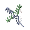
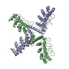
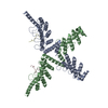
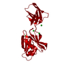
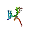
Crystal structure of mouse Tankyrase/human TRF1 complex
Components
Tankyrase-1
Telomeric repeat-binding factor 1
Keywords
TRANSFERASE / SIGNALING PROTEIN / Tankyrase / TRF1 / telomere / TRANSFERASE - SIGNALING PROTEIN complex
Function / homology
Function and homology information
TCF dependent signaling in response to WNT / Degradation of AXIN / negative regulation of maintenance of mitotic sister chromatid cohesion, telomeric / Regulation of PTEN stability and activity / positive regulation of shelterin complex assembly / negative regulation of establishment of protein localization to telomere / negative regulation of establishment of RNA localization to telomere / negative regulation of establishment of protein-containing complex localization to telomere / negative regulation of telomere maintenance via semi-conservative replication / negative regulation of telomeric D-loop disassembly ...TCF dependent signaling in response to WNT / Degradation of AXIN / negative regulation of maintenance of mitotic sister chromatid cohesion, telomeric / Regulation of PTEN stability and activity / positive regulation of shelterin complex assembly / negative regulation of establishment of protein localization to telomere / negative regulation of establishment of RNA localization to telomere / negative regulation of establishment of protein-containing complex localization to telomere / negative regulation of telomere maintenance via semi-conservative replication / negative regulation of telomeric D-loop disassembly / telomerase inhibitor activity / positive regulation of telomere maintenance via telomere lengthening / meiotic telomere clustering / regulation of telomere maintenance via telomerase / telomeric D-loop disassembly / shelterin complex / t-circle formation / Telomere C-strand synthesis initiation / double-stranded telomeric DNA binding / Ub-specific processing proteases / ankyrin repeat binding / positive regulation of telomere capping / Telomere C-strand (Lagging Strand) Synthesis / nuclear telomere cap complex / G-rich strand telomeric DNA binding / telomere capping / Polymerase switching on the C-strand of the telomere / Processive synthesis on the C-strand of the telomere / Removal of the Flap Intermediate from the C-strand / NAD+ ADP-ribosyltransferase / protein auto-ADP-ribosylation / DNA binding, bending / protein localization to chromosome, telomeric region / negative regulation of telomere maintenance via telomere lengthening / NAD+-protein-aspartate ADP-ribosyltransferase activity / protein poly-ADP-ribosylation / NAD+-protein-glutamate ADP-ribosyltransferase activity / mitotic spindle pole / negative regulation of DNA replication / telomeric DNA binding / NAD+-protein mono-ADP-ribosyltransferase activity / negative regulation of telomere maintenance via telomerase / positive regulation of telomere maintenance / Transferases; Glycosyltransferases; Pentosyltransferases / NAD+ poly-ADP-ribosyltransferase activity / Telomere Extension By Telomerase / mRNA transport / nuclear pore / telomere maintenance via telomerase / positive regulation of telomere maintenance via telomerase / Packaging Of Telomere Ends / nucleotidyltransferase activity / Recognition and association of DNA glycosylase with site containing an affected purine / Cleavage of the damaged purine / Recognition and association of DNA glycosylase with site containing an affected pyrimidine / Cleavage of the damaged pyrimidine / telomere maintenance / Inhibition of DNA recombination at telomere / Meiotic synapsis / DNA Damage/Telomere Stress Induced Senescence / spindle / fibrillar center / Wnt signaling pathway / protein polyubiquitination / positive regulation of canonical Wnt signaling pathway / protein transport / histone binding / nuclear membrane / microtubule binding / chromosome, telomeric region / nuclear body / response to xenobiotic stimulus / Golgi membrane / cell division / centrosome / nucleolus / Golgi apparatus / protein homodimerization activity / positive regulation of transcription by RNA polymerase II / DNA binding / zinc ion binding / nucleoplasm / identical protein binding / nucleus / cytoplasm / cytosol Similarity search - Function
Resolution: 2.2→79 Å / Cor.coef. Fo:Fc: 0.961 / Cor.coef. Fo:Fc free: 0.947 / SU B: 12.011 / SU ML: 0.146 / Cross valid method: THROUGHOUT / σ(F): 0 / ESU R: 0.199 / ESU R Free: 0.171 / Stereochemistry target values: MAXIMUM LIKELIHOOD Details: HYDROGENS HAVE BEEN ADDED IN THE RIDING POSITIONS U VALUES : WITH TLS ADDED
Rfactor
Num. reflection
% reflection
Selection details
Rfree
0.229
2425
5 %
RANDOM
Rwork
0.1984
-
-
-
obs
0.1999
45650
97.73 %
-
Solvent computation
Ion probe radii: 0.8 Å / Shrinkage radii: 0.8 Å / VDW probe radii: 1.2 Å / Solvent model: MASK
In the structure databanks used in Yorodumi, some data are registered as the other names, "COVID-19 virus" and "2019-nCoV". Here are the details of the virus and the list of structure data.
Jan 31, 2019. EMDB accession codes are about to change! (news from PDBe EMDB page)
EMDB accession codes are about to change! (news from PDBe EMDB page)
The allocation of 4 digits for EMDB accession codes will soon come to an end. Whilst these codes will remain in use, new EMDB accession codes will include an additional digit and will expand incrementally as the available range of codes is exhausted. The current 4-digit format prefixed with “EMD-” (i.e. EMD-XXXX) will advance to a 5-digit format (i.e. EMD-XXXXX), and so on. It is currently estimated that the 4-digit codes will be depleted around Spring 2019, at which point the 5-digit format will come into force.
The EM Navigator/Yorodumi systems omit the EMD- prefix.
Related info.:Q: What is EMD? / ID/Accession-code notation in Yorodumi/EM Navigator
Yorodumi is a browser for structure data from EMDB, PDB, SASBDB, etc.
This page is also the successor to EM Navigator detail page, and also detail information page/front-end page for Omokage search.
The word "yorodu" (or yorozu) is an old Japanese word meaning "ten thousand". "mi" (miru) is to see.
Related info.:EMDB / PDB / SASBDB / Comparison of 3 databanks / Yorodumi Search / Aug 31, 2016. New EM Navigator & Yorodumi / Yorodumi Papers / Jmol/JSmol / Function and homology information / Changes in new EM Navigator and Yorodumi
 Movie
Movie Controller


 Open data
 Basic information
 Components
 Keywords
 Function and homology information
 Homo sapiens (human)
 X-RAY DIFFRACTION /
  Authors
 Citation
 Structure visualization
 Downloads & links
 Other downloads

 PDBj
PDBj


 Assembly

 Mass: 18.015 Da / Num. of mol.: 106 / Source method: isolated from a natural source / Formula: H2O
 Sample preparation
 / Beamline: BL18U1 / Wavelength: 0.978 Å
 Processing