- PDB-5g0n: Structure of rat neuronal nitric oxide synthase D597N mutant heme... -
+
Open data
ID or keywords:
Loading...
-
Basic information
Entry
Database: PDB / ID: 5g0n
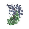
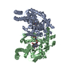
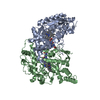
Title
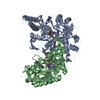
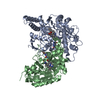
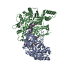
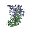
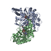
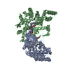
Structure of rat neuronal nitric oxide synthase D597N mutant heme domain in complex with N1-(5-(2-(6-AMINO-4-METHYLPYRIDIN-2-YL)ETHYL) PYRIDIN-3-YL)-N1,N2-DIMETHYLETHANE-1,2-DIAMINE
Nitric oxide stimulates guanylate cyclase / negative regulation of hepatic stellate cell contraction / synaptic signaling by nitric oxide / negative regulation of iron ion transmembrane transport / ROS and RNS production in phagocytes / azurophil granule / negative regulation of vasoconstriction / retrograde trans-synaptic signaling by nitric oxide / Ion homeostasis / positive regulation of sodium ion transmembrane transport ...Nitric oxide stimulates guanylate cyclase / negative regulation of hepatic stellate cell contraction / synaptic signaling by nitric oxide / negative regulation of iron ion transmembrane transport / ROS and RNS production in phagocytes / azurophil granule / negative regulation of vasoconstriction / retrograde trans-synaptic signaling by nitric oxide / Ion homeostasis / positive regulation of sodium ion transmembrane transport / positive regulation of adenylate cyclase-activating G protein-coupled receptor signaling pathway / response to nitric oxide / postsynaptic specialization, intracellular component / nitric oxide metabolic process / negative regulation of cytosolic calcium ion concentration / peptidyl-cysteine S-nitrosylation / behavioral response to cocaine / postsynaptic density, intracellular component / cadmium ion binding / positive regulation of the force of heart contraction / negative regulation of potassium ion transport / calyx of Held / negative regulation of calcium ion transport / negative regulation of serotonin uptake / sodium channel regulator activity / regulation of neurogenesis / striated muscle contraction / negative regulation of insulin secretion / response to vitamin E / nitric-oxide synthase (NADPH) / regulation of postsynaptic membrane potential / multicellular organismal response to stress / xenobiotic catabolic process / nitric-oxide synthase activity / negative regulation of peptidyl-serine phosphorylation / nitric oxide mediated signal transduction / arginine catabolic process / NADPH binding / regulation of sodium ion transport / response to organonitrogen compound / cellular response to epinephrine stimulus / sarcoplasmic reticulum membrane / T-tubule / photoreceptor inner segment / nitric oxide biosynthetic process / negative regulation of blood pressure / response to hormone / response to nutrient levels / response to activity / response to nicotine / sarcoplasmic reticulum / muscle contraction / secretory granule / female pregnancy / positive regulation of long-term synaptic potentiation / cell periphery / establishment of localization in cell / phosphoprotein binding / response to lead ion / sarcolemma / establishment of protein localization / potassium ion transport / response to organic cyclic compound / response to peptide hormone / cellular response to growth factor stimulus / Z disc / vasodilation / cellular response to mechanical stimulus / response to estrogen / calcium-dependent protein binding / calcium ion transport / FMN binding / flavin adenine dinucleotide binding / positive regulation of peptidyl-serine phosphorylation / NADP binding / ATPase binding / response to heat / scaffold protein binding / response to ethanol / nuclear membrane / negative regulation of neuron apoptotic process / mitochondrial outer membrane / transmembrane transporter binding / response to lipopolysaccharide / dendritic spine / postsynaptic density / response to hypoxia / cytoskeleton / calmodulin binding / membrane raft / negative regulation of cell population proliferation / dendrite / glutamatergic synapse / synapse / heme binding / negative regulation of apoptotic process / perinuclear region of cytoplasm / positive regulation of DNA-templated transcription / enzyme binding / positive regulation of transcription by RNA polymerase II Similarity search - Function
NITRICOXIDESYNTHASE, BRAIN / / BNOS / CONSTITUTIVE NOS / NC-NOS / NOS TYPE I / NEURONAL NOS / N-NOS / NNOS / PEPTIDYL-CYSTEINE S- ...BNOS / CONSTITUTIVE NOS / NC-NOS / NOS TYPE I / NEURONAL NOS / N-NOS / NNOS / PEPTIDYL-CYSTEINE S-NITROSYLASE NOS1 / NEURONAL NITRIC OXIDE SYNTHASE
Mass: 48811.543 Da / Num. of mol.: 2 / Fragment: HEME DOMAIN / Mutation: YES Source method: isolated from a genetically manipulated source Source: (gene. exp.) RATTUS NORVEGICUS (Norway rat) / Production host: ESCHERICHIA COLI (E. coli) / Strain (production host): BL21(DE3) / References: UniProt: P29476, nitric-oxide synthase (NADPH)
-
Non-polymers , 6 types, 507 molecules
#2: Chemical
ChemComp-HEM / PROTOPORPHYRINIXCONTAININGFE / HEME / Heme B
Mass: 616.487 Da / Num. of mol.: 2 / Source method: obtained synthetically / Formula: C34H32FeN4O4
Monochromator: GRAPHITE / Protocol: SINGLE WAVELENGTH / Monochromatic (M) / Laue (L): M / Scattering type: x-ray
Radiation wavelength
Wavelength: 1.127 Å / Relative weight: 1
Reflection
Resolution: 1.94→50 Å / Num. obs: 70703 / % possible obs: 99.6 % / Observed criterion σ(I): -3 / Redundancy: 5 % / Biso Wilson estimate: 28.95 Å2 / Rmerge(I) obs: 0.11 / Net I/σ(I): 9.1
Reflection shell
Resolution: 1.94→2 Å / Redundancy: 4.2 % / Rmerge(I) obs: 1.23 / Mean I/σ(I) obs: 1.1 / % possible all: 93.4
-
Processing
Software
Name
Version
Classification
PHENIX
(PHENIX.REFINE)
refinement
XDS
datareduction
Aimless
datascaling
REFMAC
phasing
Refinement
Method to determine structure: OTHER Starting model: NONE Resolution: 1.936→38.828 Å / SU ML: 0.28 / σ(F): 1.13 / Phase error: 24.76 / Stereochemistry target values: ML Details: RESIDUES 339-349 IN CHAIN A AND 339-347 IN CHAIN B ARE DISORDERED.
Rfactor
Num. reflection
% reflection
Rfree
0.2157
6676
5 %
Rwork
0.1753
-
-
obs
0.1773
134166
99.23 %
Solvent computation
Shrinkage radii: 0.9 Å / VDW probe radii: 1.11 Å / Solvent model: FLAT BULK SOLVENT MODEL
Refinement step
Cycle: LAST / Resolution: 1.936→38.828 Å
Protein
Nucleic acid
Ligand
Solvent
Total
Num. atoms
6659
0
173
498
7330
Refine LS restraints
Refine-ID
Type
Dev ideal
Number
X-RAY DIFFRACTION
f_bond_d
0.007
7069
X-RAY DIFFRACTION
f_angle_d
1.141
9618
X-RAY DIFFRACTION
f_dihedral_angle_d
15.182
2574
X-RAY DIFFRACTION
f_chiral_restr
0.074
994
X-RAY DIFFRACTION
f_plane_restr
0.005
1217
LS refinement shell
Resolution (Å)
Rfactor Rfree
Num. reflection Rfree
Rfactor Rwork
Num. reflection Rwork
Refine-ID
% reflection obs (%)
1.9358-1.9577
0.4059
190
0.3743
3487
X-RAY DIFFRACTION
82
1.9577-1.9808
0.3815
248
0.3423
4204
X-RAY DIFFRACTION
99
1.9808-2.0049
0.4145
207
0.3191
4303
X-RAY DIFFRACTION
100
2.0049-2.0303
0.2804
203
0.287
4250
X-RAY DIFFRACTION
100
2.0303-2.057
0.2968
226
0.2794
4259
X-RAY DIFFRACTION
100
2.057-2.0852
0.3465
238
0.2741
4284
X-RAY DIFFRACTION
100
2.0852-2.115
0.3446
177
0.2695
4300
X-RAY DIFFRACTION
100
2.115-2.1466
0.2744
203
0.2469
4321
X-RAY DIFFRACTION
100
2.1466-2.1801
0.3308
198
0.2535
4294
X-RAY DIFFRACTION
100
2.1801-2.2158
0.3332
228
0.2406
4290
X-RAY DIFFRACTION
100
2.2158-2.254
0.2612
238
0.2312
4307
X-RAY DIFFRACTION
100
2.254-2.295
0.2651
259
0.2175
4193
X-RAY DIFFRACTION
100
2.295-2.3392
0.2338
244
0.2097
4257
X-RAY DIFFRACTION
100
2.3392-2.3869
0.2485
220
0.2025
4303
X-RAY DIFFRACTION
100
2.3869-2.4388
0.264
195
0.2077
4290
X-RAY DIFFRACTION
100
2.4388-2.4955
0.3117
214
0.2048
4289
X-RAY DIFFRACTION
100
2.4955-2.5579
0.2586
236
0.1935
4258
X-RAY DIFFRACTION
100
2.5579-2.6271
0.228
226
0.1839
4274
X-RAY DIFFRACTION
100
2.6271-2.7043
0.2363
210
0.172
4316
X-RAY DIFFRACTION
100
2.7043-2.7916
0.2056
204
0.1649
4274
X-RAY DIFFRACTION
100
2.7916-2.8913
0.2029
241
0.1682
4311
X-RAY DIFFRACTION
100
2.8913-3.0071
0.2065
231
0.1707
4218
X-RAY DIFFRACTION
100
3.0071-3.1439
0.2573
241
0.175
4286
X-RAY DIFFRACTION
100
3.1439-3.3095
0.2146
205
0.1645
4279
X-RAY DIFFRACTION
100
3.3095-3.5168
0.1736
214
0.1511
4306
X-RAY DIFFRACTION
100
3.5168-3.7881
0.2025
259
0.1337
4264
X-RAY DIFFRACTION
100
3.7881-4.1689
0.1717
213
0.1206
4266
X-RAY DIFFRACTION
100
4.1689-4.7712
0.1382
244
0.1173
4282
X-RAY DIFFRACTION
100
4.7712-6.0076
0.1829
253
0.1361
4223
X-RAY DIFFRACTION
100
6.0076-38.8355
0.1381
211
0.1523
4302
X-RAY DIFFRACTION
100
Refinement TLS params.
Method: refined / Refine-ID: X-RAY DIFFRACTION
ID
L11 (°2)
L12 (°2)
L13 (°2)
L22 (°2)
L23 (°2)
L33 (°2)
S11 (Å °)
S12 (Å °)
S13 (Å °)
S21 (Å °)
S22 (Å °)
S23 (Å °)
S31 (Å °)
S32 (Å °)
S33 (Å °)
T11 (Å2)
T12 (Å2)
T13 (Å2)
T22 (Å2)
T23 (Å2)
T33 (Å2)
Origin x (Å)
Origin y (Å)
Origin z (Å)
1
0.612
-0.0552
-0.3669
0.8352
-0.2531
3.9867
-0.0595
0.1107
-0.0223
0.0121
-0.0244
0.032
-0.0228
-0.2997
0.0572
0.1632
-0.0225
-0.0006
0.2067
-0.0153
0.1905
11.3816
4.8823
22.4398
2
0.661
-0.1217
-0.1222
0.8418
0.3578
2.0437
-0.0173
-0.0017
0.0496
-0.0839
-0.035
-0.0175
0.0118
0.0742
0.0497
0.1347
0.0043
0.0162
0.1449
0.013
0.1872
12.094
4.8045
59.7331
Refinement TLS group
ID
Refine-ID
Refine TLS-ID
Selection details
1
X-RAY DIFFRACTION
1
(CHAINAANDRESID299:716)
2
X-RAY DIFFRACTION
2
(CHAINBANDRESID299:718)
+
About Yorodumi
-
News
-
Feb 9, 2022. New format data for meta-information of EMDB entries
New format data for meta-information of EMDB entries
Version 3 of the EMDB header file is now the official format.
The previous official version 1.9 will be removed from the archive.
In the structure databanks used in Yorodumi, some data are registered as the other names, "COVID-19 virus" and "2019-nCoV". Here are the details of the virus and the list of structure data.
Jan 31, 2019. EMDB accession codes are about to change! (news from PDBe EMDB page)
EMDB accession codes are about to change! (news from PDBe EMDB page)
The allocation of 4 digits for EMDB accession codes will soon come to an end. Whilst these codes will remain in use, new EMDB accession codes will include an additional digit and will expand incrementally as the available range of codes is exhausted. The current 4-digit format prefixed with “EMD-” (i.e. EMD-XXXX) will advance to a 5-digit format (i.e. EMD-XXXXX), and so on. It is currently estimated that the 4-digit codes will be depleted around Spring 2019, at which point the 5-digit format will come into force.
The EM Navigator/Yorodumi systems omit the EMD- prefix.
Related info.:Q: What is EMD? / ID/Accession-code notation in Yorodumi/EM Navigator
Yorodumi is a browser for structure data from EMDB, PDB, SASBDB, etc.
This page is also the successor to EM Navigator detail page, and also detail information page/front-end page for Omokage search.
The word "yorodu" (or yorozu) is an old Japanese word meaning "ten thousand". "mi" (miru) is to see.
Related info.:EMDB / PDB / SASBDB / Comparison of 3 databanks / Yorodumi Search / Aug 31, 2016. New EM Navigator & Yorodumi / Yorodumi Papers / Jmol/JSmol / Function and homology information / Changes in new EM Navigator and Yorodumi
 Movie
Movie Controller

 Yorodumi
 Open data
 Basic information
 Components
 Keywords
 Function and homology information
 Authors
 Citation
 Structure visualization
 Downloads & links
 Other downloads
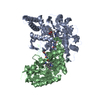


 PDBj
PDBj


 Assembly
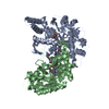


 Mass: 616.487 Da / Num. of mol.: 2 / Source method: obtained synthetically / Formula: C34H32FeN4O4
 Mass: 241.247 Da / Num. of mol.: 2 / Source method: obtained synthetically / Formula: C9H15N5O3 / Comment: neurotransmitter*YM
 Mass: 299.414 Da / Num. of mol.: 2 / Source method: obtained synthetically / Formula: C17H25N5
 Mass: 59.044 Da / Num. of mol.: 2 / Source method: obtained synthetically / Formula: C2H3O2
 Mass: 65.409 Da / Num. of mol.: 1 / Source method: obtained synthetically / Formula: Zn
 Sample preparation
 / Beamline: BL7-1 / Wavelength: 1.127
  Processing