 Movie
Movie Controller

[English] 日本語
 Yorodumi
- PDB-5dsd: The crystal structure of the C-terminal domain of Ebola (Bundibug... -
+ Open data

- Basic information
| Entry | Database: PDB / ID: 5dsd | |||||||||
|---|---|---|---|---|---|---|---|---|---|---|
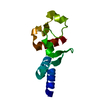
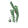
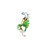
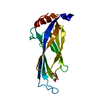
| Title | The crystal structure of the C-terminal domain of Ebola (Bundibugyo) nucleoprotein | |||||||||
 Components Components | Nucleoprotein | |||||||||
 Keywords Keywords | VIRAL PROTEIN / Filoviridae | |||||||||
| Function / homology |  Function and homology information Function and homology informationviral RNA genome packaging / helical viral capsid / viral nucleocapsid / host cell cytoplasm / ribonucleoprotein complex / RNA binding Similarity search - Function | |||||||||
| Biological species |  Bundibugyo virus Bundibugyo virus | |||||||||
| Method |  X-RAY DIFFRACTION / SYNCHROTRON / MOLECULAR REPLACEMENT / Resolution: 2.31 Å X-RAY DIFFRACTION / SYNCHROTRON / MOLECULAR REPLACEMENT / Resolution: 2.31 Å | |||||||||
 Authors Authors | Baker, L. / Handing, K.B. / Utepbergenov, D. / Derewenda, U. / Derewenda, Z.S. | |||||||||
| Funding support |  United States, 1items United States, 1items
| |||||||||
 Citation Citation | Journal: Acta Crystallogr D Struct Biol / Year: 2016 Title: Molecular architecture of the nucleoprotein C-terminal domain from the Ebola and Marburg viruses. Authors: Baker, L.E. / Ellena, J.F. / Handing, K.B. / Derewenda, U. / Utepbergenov, D. / Engel, D.A. / Derewenda, Z.S. #1: Journal: Acta Crystallogr. D Biol. Crystallogr. / Year: 2014Title: The structure of the C-terminal domain of the Zaire ebolavirus nucleoprotein. Authors: Dziubanska, P.J. / Derewenda, U. / Ellena, J.F. / Engel, D.A. / Derewenda, Z.S. | |||||||||
| History |
|
- Structure visualization
| Structure viewer | Molecule: MolmilJmol/JSmol |
|---|
- Downloads & links
-Download
| PDBx/mmCIF format | 5dsd.cif.gz | 65 KB | Display | PDBx/mmCIF format |
|---|---|---|---|---|
| PDB format | pdb5dsd.ent.gz | 46.7 KB | Display | PDB format |
| PDBx/mmJSON format | 5dsd.json.gz | Tree view | PDBx/mmJSON format | |
| Others |  Other downloads Other downloads |
-Validation report
| Arichive directory | https://data.pdbj.org/pub/pdb/validation_reports/ds/5dsdftp://data.pdbj.org/pub/pdb/validation_reports/ds/5dsd | HTTPS FTP |
|---|
-Related structure data
| Related structure data | 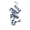 5e2xC  4qb0S S: Starting model for refinement C: citing same article ( |
|---|---|
| Similar structure data |


-Links
 PDBj
PDBj
- Assembly
| Deposited unit | 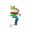
| ||||||||
|---|---|---|---|---|---|---|---|---|---|
| 1 |
| ||||||||
| 2 | 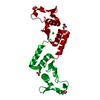
| ||||||||
| 3 | 
| ||||||||
| Unit cell |
|
-Components
| #1: Protein | Mass: 12267.726 Da / Num. of mol.: 1 / Fragment: C-terminal domain (UNP residues 641-739) Source method: isolated from a genetically manipulated source Source: (gene. exp.) Bundibugyo virus / Gene: NP, DH33_45404gpNP / Plasmid: Parallel1-MBPHis / Production host:  | ||||
|---|---|---|---|---|---|
| #2: Chemical |   Mass: 92.094 Da / Num. of mol.: 2 / Source method: obtained synthetically / Formula: C3H8O3 Mass: 92.094 Da / Num. of mol.: 2 / Source method: obtained synthetically / Formula: C3H8O3#3: Chemical | ChemComp-CL / |   Mass: 35.453 Da / Num. of mol.: 1 / Source method: obtained synthetically / Formula: Cl Mass: 35.453 Da / Num. of mol.: 1 / Source method: obtained synthetically / Formula: Cl#4: Water | ChemComp-HOH / |  Mass: 18.015 Da / Num. of mol.: 112 / Source method: isolated from a natural source / Formula: H2O Mass: 18.015 Da / Num. of mol.: 112 / Source method: isolated from a natural source / Formula: H2O |
-Experimental details
-Experiment
| Experiment | Method: X-RAY DIFFRACTION / Number of used crystals: 1 |
|---|
- Sample preparation
| Crystal | Density Matthews: 5.94 Å3/Da / Density % sol: 79 % |
|---|---|
| Crystal grow | Temperature: 293 K / Method: vapor diffusion, sitting drop / pH: 8.5 Details: Crystallization solution: 1.33 M LiSO4, 0.1 M Tris pH 8.5. 1:1 ratio (250 nL: 250 nL) of precipitant to protein (at a concentration of 13.9 mg/ml). 60 mcL reservoir. Protein purification ...Details: Crystallization solution: 1.33 M LiSO4, 0.1 M Tris pH 8.5. 1:1 ratio (250 nL: 250 nL) of precipitant to protein (at a concentration of 13.9 mg/ml). 60 mcL reservoir. Protein purification buffer 50mM Tris-HCl, 150 mM NaCl; pH 7.5 |
-Data collection
| Diffraction | Mean temperature: 100 K |
|---|---|
| Diffraction source | Source: SYNCHROTRON / Site: APS / Beamline: 21-ID-D / Wavelength: 0.99 Å |
| Detector | Type: RAYONIX MX300HE / Detector: CCD / Date: Aug 14, 2014 |
| Diffraction measurement | Details: 0.50 degrees, -1.0 sec, detector distance 350.62 mm Method: \w scans |
| Radiation | Monochromator: Si(111) / Protocol: SINGLE WAVELENGTH / Monochromatic (M) / Laue (L): M / Scattering type: x-ray |
| Radiation wavelength | Wavelength: 0.99 Å / Relative weight: 1 |
| Reflection | Av R equivalents: 0.084 / Number: 184210 |
| Reflection | Resolution: 2.3→80 Å / Num. obs: 12398 / % possible obs: 94.3 % / Redundancy: 14.8 % / Rmerge(I) obs: 0.084 / Net I/av σ(I): 31.34 / Net I/σ(I): 20.3 |
| Reflection shell | Resolution: 2.3→2.38 Å / Redundancy: 7.1 % / Rmerge(I) obs: 0.432 / Mean I/σ(I) obs: 1.9 / Rsym value: 0.432 / % possible all: 64.7 |
| Cell measurement | Reflection used: 184210 |
- Processing
| Software |
| |||||||||||||||||||||||||||||||||||||||||||||||||||||||||||||||||||||||||||||||||||||||||||||||||||||||||||||||||||||||||||||||||||||||||||||||||||||||||||||||||||||||||||||||
|---|---|---|---|---|---|---|---|---|---|---|---|---|---|---|---|---|---|---|---|---|---|---|---|---|---|---|---|---|---|---|---|---|---|---|---|---|---|---|---|---|---|---|---|---|---|---|---|---|---|---|---|---|---|---|---|---|---|---|---|---|---|---|---|---|---|---|---|---|---|---|---|---|---|---|---|---|---|---|---|---|---|---|---|---|---|---|---|---|---|---|---|---|---|---|---|---|---|---|---|---|---|---|---|---|---|---|---|---|---|---|---|---|---|---|---|---|---|---|---|---|---|---|---|---|---|---|---|---|---|---|---|---|---|---|---|---|---|---|---|---|---|---|---|---|---|---|---|---|---|---|---|---|---|---|---|---|---|---|---|---|---|---|---|---|---|---|---|---|---|---|---|---|---|---|---|---|
| Refinement | Method to determine structure: MOLECULAR REPLACEMENT Starting model: 4QB0 Resolution: 2.31→80 Å / Cor.coef. Fo:Fc: 0.97 / Cor.coef. Fo:Fc free: 0.964 / WRfactor Rfree: 0.2071 / WRfactor Rwork: 0.1698 / FOM work R set: 0.8163 / SU B: 9.71 / SU ML: 0.117 / SU R Cruickshank DPI: 0.1436 / SU Rfree: 0.1412 / Cross valid method: THROUGHOUT / σ(F): 0 / ESU R: 0.144 / ESU R Free: 0.141 / Stereochemistry target values: MAXIMUM LIKELIHOOD Details: U VALUES : WITH TLS ADDED HYDROGENS HAVE BEEN ADDED IN THE RIDING POSITIONS
| |||||||||||||||||||||||||||||||||||||||||||||||||||||||||||||||||||||||||||||||||||||||||||||||||||||||||||||||||||||||||||||||||||||||||||||||||||||||||||||||||||||||||||||||
| Solvent computation | Ion probe radii: 0.8 Å / Shrinkage radii: 0.8 Å / VDW probe radii: 1.2 Å / Solvent model: BABINET MODEL WITH MASK | |||||||||||||||||||||||||||||||||||||||||||||||||||||||||||||||||||||||||||||||||||||||||||||||||||||||||||||||||||||||||||||||||||||||||||||||||||||||||||||||||||||||||||||||
| Displacement parameters | Biso max: 243.89 Å2 / Biso mean: 81.78 Å2 / Biso min: 46.22 Å2
| |||||||||||||||||||||||||||||||||||||||||||||||||||||||||||||||||||||||||||||||||||||||||||||||||||||||||||||||||||||||||||||||||||||||||||||||||||||||||||||||||||||||||||||||
| Refinement step | Cycle: final / Resolution: 2.31→80 Å
| |||||||||||||||||||||||||||||||||||||||||||||||||||||||||||||||||||||||||||||||||||||||||||||||||||||||||||||||||||||||||||||||||||||||||||||||||||||||||||||||||||||||||||||||
| Refine LS restraints |
| |||||||||||||||||||||||||||||||||||||||||||||||||||||||||||||||||||||||||||||||||||||||||||||||||||||||||||||||||||||||||||||||||||||||||||||||||||||||||||||||||||||||||||||||
| LS refinement shell | Resolution: 2.312→2.372 Å / Total num. of bins used: 20
| |||||||||||||||||||||||||||||||||||||||||||||||||||||||||||||||||||||||||||||||||||||||||||||||||||||||||||||||||||||||||||||||||||||||||||||||||||||||||||||||||||||||||||||||
| Refinement TLS params. | Method: refined / Refine-ID: X-RAY DIFFRACTION
| |||||||||||||||||||||||||||||||||||||||||||||||||||||||||||||||||||||||||||||||||||||||||||||||||||||||||||||||||||||||||||||||||||||||||||||||||||||||||||||||||||||||||||||||
| Refinement TLS group |
|
