 Movie
Movie Controller

[English] 日本語
 Yorodumi
- PDB-5d7b: Trigonal Crystal Structure of an acetylester hydrolase from Coryn... -
+ Open data

- Basic information
| Entry | Database: PDB / ID: 5d7b | ||||||
|---|---|---|---|---|---|---|---|
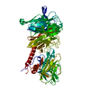
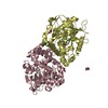
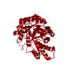
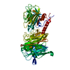
| Title | Trigonal Crystal Structure of an acetylester hydrolase from Corynebacterium glutamicum | ||||||
 Components Components | Homoserine O-acetyltransferase | ||||||
 Keywords Keywords | HYDROLASE / Acetylester hydrolase / Alpha/Beta-Hydrolase | ||||||
| Function / homology |  Function and homology information Function and homology informationhomoserine O-acetyltransferase activity / Hydrolases; Acting on ester bonds; Carboxylic-ester hydrolases / biosynthetic process / hydrolase activity Similarity search - Function | ||||||
| Biological species |  Corynebacterium glutamicum (bacteria) Corynebacterium glutamicum (bacteria) | ||||||
| Method |  X-RAY DIFFRACTION / SYNCHROTRON / MOLECULAR REPLACEMENT / Resolution: 3.2 Å X-RAY DIFFRACTION / SYNCHROTRON / MOLECULAR REPLACEMENT / Resolution: 3.2 Å | ||||||
 Authors Authors | Niefind, K. / Toelzer, C. / Pal, S. / Altenbuchner, J. / Watzlawick, H. | ||||||
| Funding support |  Germany, 1items Germany, 1items
| ||||||
 Citation Citation | Journal: Febs Lett. / Year: 2016 Title: A novel esterase subfamily with alpha / beta-hydrolase fold suggested by structures of two bacterial enzymes homologous to l-homoserine O-acetyl transferases. Authors: Tolzer, C. / Pal, S. / Watzlawick, H. / Altenbuchner, J. / Niefind, K. | ||||||
| History |
|
- Structure visualization
| Structure viewer | Molecule: MolmilJmol/JSmol |
|---|
- Downloads & links
-Download
| PDBx/mmCIF format | 5d7b.cif.gz | 290.4 KB | Display | PDBx/mmCIF format |
|---|---|---|---|---|
| PDB format | pdb5d7b.ent.gz | 240.9 KB | Display | PDB format |
| PDBx/mmJSON format | 5d7b.json.gz | Tree view | PDBx/mmJSON format | |
| Others |  Other downloads Other downloads |
-Validation report
| Arichive directory | https://data.pdbj.org/pub/pdb/validation_reports/d7/5d7bftp://data.pdbj.org/pub/pdb/validation_reports/d7/5d7b | HTTPS FTP |
|---|
-Related structure data
| Related structure data |  5d6oC  5e4yC  5efzC  2b61S S: Starting model for refinement C: citing same article ( |
|---|---|
| Similar structure data |


-Links
 PDBj
PDBj


- Assembly
| Deposited unit | 
| ||||||||||||||||||
|---|---|---|---|---|---|---|---|---|---|---|---|---|---|---|---|---|---|---|---|
| 1 |
| ||||||||||||||||||
| Unit cell |
| ||||||||||||||||||
| Components on special symmetry positions |
| ||||||||||||||||||
| Noncrystallographic symmetry (NCS) | NCS domain:
NCS domain segments:
|
-Components
| #1: Protein | Mass: 38870.207 Da / Num. of mol.: 2 Source method: isolated from a genetically manipulated source Source: (gene. exp.) Corynebacterium glutamicum (bacteria) / Gene: cg0961, Cgl0839 / Plasmid: pHWG771 / Production host: References: UniProt: Q8NS43, Transferases; Acyltransferases; Transferring groups other than aminoacyl groups, homoserine O-acetyltransferase #2: Chemical | ChemComp-GOL /   Mass: 92.094 Da / Num. of mol.: 15 / Source method: obtained synthetically / Formula: C3H8O3 Mass: 92.094 Da / Num. of mol.: 15 / Source method: obtained synthetically / Formula: C3H8O3#3: Water | ChemComp-HOH / |  Mass: 18.015 Da / Num. of mol.: 17 / Source method: isolated from a natural source / Formula: H2O Mass: 18.015 Da / Num. of mol.: 17 / Source method: isolated from a natural source / Formula: H2O |
|---|
-Experimental details
-Experiment
| Experiment | Method: X-RAY DIFFRACTION |
|---|
- Sample preparation
| Crystal | Density Matthews: 7.52 Å3/Da / Density % sol: 83.7 % |
|---|---|
| Crystal grow | Temperature: 293 K / Method: vapor diffusion, sitting drop / pH: 8.5 Details: Reservoir: 25.5 %(w/v) polyethylenglycol 4000, 15 %(v/v) glycerol, 0.17 M Lithium sulfate, 85 mM Tris/HCl, pH 8.5 Drop: 0.4 microliter reservoir solution plut 0.8 microliter protein solution ...Details: Reservoir: 25.5 %(w/v) polyethylenglycol 4000, 15 %(v/v) glycerol, 0.17 M Lithium sulfate, 85 mM Tris/HCl, pH 8.5 Drop: 0.4 microliter reservoir solution plut 0.8 microliter protein solution with 5 mg/ml protein concentration |
-Data collection
| Diffraction | Mean temperature: 100 K |
|---|---|
| Diffraction source | Source: SYNCHROTRON / Site: BESSY / Beamline: 14.1 / Wavelength: 0.91841 Å |
| Detector | Type: MARMOSAIC 225 mm CCD / Detector: CCD / Date: Jul 26, 2008 |
| Radiation | Protocol: SINGLE WAVELENGTH / Monochromatic (M) / Laue (L): M / Scattering type: x-ray |
| Radiation wavelength | Wavelength: 0.91841 Å / Relative weight: 1 |
| Reflection | Resolution: 3.2→48.6 Å / Num. obs: 39336 / % possible obs: 99.93 % / Redundancy: 9.5 % / Rsym value: 0.245 / Net I/σ(I): 24.66 |
| Reflection shell | Resolution: 3.2→3.314 Å / Redundancy: 9.7 % / Rmerge(I) obs: 1.01 / Mean I/σ(I) obs: 2.66 / % possible all: 99.95 |
- Processing
| Software |
| ||||||||||||||||||||||||||||||||||||||||||||||||||||||||||||||||||||||||
|---|---|---|---|---|---|---|---|---|---|---|---|---|---|---|---|---|---|---|---|---|---|---|---|---|---|---|---|---|---|---|---|---|---|---|---|---|---|---|---|---|---|---|---|---|---|---|---|---|---|---|---|---|---|---|---|---|---|---|---|---|---|---|---|---|---|---|---|---|---|---|---|---|---|
| Refinement | Method to determine structure: MOLECULAR REPLACEMENT Starting model: 2B61 Resolution: 3.2→48.529 Å / SU ML: 0.26 / Cross valid method: FREE R-VALUE / σ(F): 1.35 / Phase error: 19.73 / Stereochemistry target values: ML
| ||||||||||||||||||||||||||||||||||||||||||||||||||||||||||||||||||||||||
| Solvent computation | Shrinkage radii: 0.9 Å / VDW probe radii: 1.11 Å / Solvent model: FLAT BULK SOLVENT MODEL | ||||||||||||||||||||||||||||||||||||||||||||||||||||||||||||||||||||||||
| Refinement step | Cycle: LAST / Resolution: 3.2→48.529 Å
| ||||||||||||||||||||||||||||||||||||||||||||||||||||||||||||||||||||||||
| Refine LS restraints |
| ||||||||||||||||||||||||||||||||||||||||||||||||||||||||||||||||||||||||
| Refine LS restraints NCS |
| ||||||||||||||||||||||||||||||||||||||||||||||||||||||||||||||||||||||||
| LS refinement shell |
| ||||||||||||||||||||||||||||||||||||||||||||||||||||||||||||||||||||||||
| Refinement TLS params. | L12: -0.8488 °2 / Method: refined / Refine-ID: X-RAY DIFFRACTION
| ||||||||||||||||||||||||||||||||||||||||||||||||||||||||||||||||||||||||
| Refinement TLS group |
|
