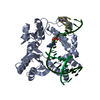
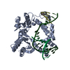
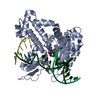
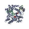
 Movie
Movie Controller


+ Open data

- Basic information
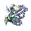
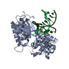
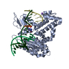
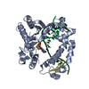
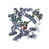
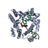
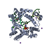
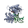
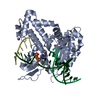
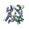
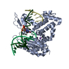
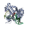
| Entry | Database: PDB / ID: 5bom | ||||||
|---|---|---|---|---|---|---|---|
| Title | DNA polymerase beta binary complex with a templating 5ClC | ||||||
 Components Components |
| ||||||
 Keywords Keywords |  TRANSFERASE / LYASE/DNA / LYASE-DNA complex TRANSFERASE / LYASE/DNA / LYASE-DNA complex | ||||||
| Function / homology |  Function and homology information Function and homology informationResolution of AP sites via the single-nucleotide replacement pathway / immunoglobulin heavy chain V-D-J recombination / Resolution of AP sites via the multiple-nucleotide patch replacement pathway / Abasic sugar-phosphate removal via the single-nucleotide replacement pathway / APEX1-Independent Resolution of AP Sites via the Single Nucleotide Replacement Pathway / Lyases; Carbon-oxygen lyases; Other carbon-oxygen lyases / homeostasis of number of cells / 5'-deoxyribose-5-phosphate lyase activity / PCNA-Dependent Long Patch Base Excision Repair / POLB-Dependent Long Patch Base Excision Repair ...Resolution of AP sites via the single-nucleotide replacement pathway / immunoglobulin heavy chain V-D-J recombination / Resolution of AP sites via the multiple-nucleotide patch replacement pathway / Abasic sugar-phosphate removal via the single-nucleotide replacement pathway / APEX1-Independent Resolution of AP Sites via the Single Nucleotide Replacement Pathway / Lyases; Carbon-oxygen lyases; Other carbon-oxygen lyases / homeostasis of number of cells / 5'-deoxyribose-5-phosphate lyase activity / PCNA-Dependent Long Patch Base Excision Repair / POLB-Dependent Long Patch Base Excision Repair / pyrimidine dimer repair / response to hyperoxia / somatic hypermutation of immunoglobulin genes / lymph node development / salivary gland morphogenesis / class I DNA-(apurinic or apyrimidinic site) endonuclease activity / base-excision repair, gap-filling / spleen development / DNA-(apurinic or apyrimidinic site) endonuclease activity / DNA-(apurinic or apyrimidinic site) lyase / spindle microtubule / response to gamma radiation / base-excision repair / DNA-templated DNA replication / double-strand break repair via nonhomologous end joining / intrinsic apoptotic signaling pathway in response to DNA damage / microtubule binding / neuron apoptotic process / response to ethanol / in utero embryonic development / microtubule / damaged DNA binding / DNA-directed DNA polymerase / DNA-directed DNA polymerase activity / lyase activity / Ub-specific processing proteases / inflammatory response / DNA repair / DNA damage response / enzyme binding / protein-containing complex / nucleoplasm / metal ion binding / nucleus / cytoplasmSimilarity search - Function | ||||||
| Biological species |  Homo sapiens (human) Homo sapiens (human)synthetic construct (others) | ||||||
| Method | X-RAY DIFFRACTION / MOLECULAR REPLACEMENT / Resolution: 2.0002 Å | ||||||
 Authors Authors | Freudenthal, B.D. / Wilson, S.H. | ||||||
| Funding support |  United States, 1items United States, 1items
| ||||||
 Citation Citation | Journal: Proc.Natl.Acad.Sci.USA / Year: 2015 Title: Intrinsic mutagenic properties of 5-chlorocytosine: A mechanistic connection between chronic inflammation and cancer. Authors: Fedeles, B.I. / Freudenthal, B.D. / Yau, E. / Singh, V. / Chang, S.C. / Li, D. / Delaney, J.C. / Wilson, S.H. / Essigmann, J.M. | ||||||
| History |
|
- Structure visualization
| Structure viewer | Molecule: MolmilJmol/JSmol |
|---|
- Downloads & links
-Download
| PDBx/mmCIF format | 5bom.cif.gz | 108.6 KB | Display | PDBx/mmCIF format |
|---|---|---|---|---|
| PDB format | pdb5bom.ent.gz | 76.1 KB | Display | PDB format |
| PDBx/mmJSON format | 5bom.json.gz | Tree view | PDBx/mmJSON format | |
| Others |  Other downloads Other downloads |
-Validation report
| Arichive directory | https://data.pdbj.org/pub/pdb/validation_reports/bo/5bomftp://data.pdbj.org/pub/pdb/validation_reports/bo/5bom | HTTPS FTP |
|---|
-Related structure data
| Related structure data |  5bolC  5bpcC 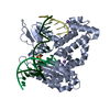 3isbS S: Starting model for refinement C: citing same article ( |
|---|---|
| Similar structure data |


-Links
 PDBj
PDBj


- Assembly
| Deposited unit | 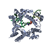
| ||||||||
|---|---|---|---|---|---|---|---|---|---|
| 1 |
| ||||||||
| Unit cell |
|
-Components
-Protein , 1 types, 1 molecules A
| #1: Protein | Mass: 38241.672 Da / Num. of mol.: 1 Source method: isolated from a genetically manipulated source Source: (gene. exp.) Homo sapiens (human) / Gene: POLB / Plasmid: pWL11 / Production host:  Escherichia coli (E. coli) Escherichia coli (E. coli)References: UniProt: P06746, DNA-directed DNA polymerase, Lyases; Carbon-oxygen lyases; Other carbon-oxygen lyases |
|---|
-DNA chain , 3 types, 3 molecules DPT
| #2: DNA chain | Mass: 1536.035 Da / Num. of mol.: 1 / Source method: obtained synthetically / Source: (synth.) synthetic construct (others) |
|---|---|
| #3: DNA chain | Mass: 3061.004 Da / Num. of mol.: 1 / Source method: obtained synthetically / Source: (synth.) synthetic construct (others) |
| #4: DNA chain | Mass: 4863.578 Da / Num. of mol.: 1 / Source method: obtained synthetically / Source: (synth.) synthetic construct (others) |
-Non-polymers , 2 types, 283 molecules 

| #5: Chemical |  Mass: 22.990 Da / Num. of mol.: 3 / Source method: obtained synthetically / Formula: Na Mass: 22.990 Da / Num. of mol.: 3 / Source method: obtained synthetically / Formula: Na#6: Water | ChemComp-HOH / | WaterMass: 18.015 Da / Num. of mol.: 280 / Source method: isolated from a natural source / Formula: H2O |
|---|
-Experimental details
-Experiment
| Experiment | Method: X-RAY DIFFRACTION / Number of used crystals: 1 |
|---|
- Sample preparation
| Crystal | Density Matthews: 2.38 Å3/Da / Density % sol: 48.52 % |
|---|---|
| Crystal grow | Temperature: 291 K / Method: vapor diffusion / pH: 7.5 Details: 50mM Imidazole, 350mM Sodium Acetate, 18% Peg3350, pH 8.0, VAPOR DIFFUSION, temperature 291K |
-Data collection
| Diffraction | Mean temperature: 100 K |
|---|---|
| Diffraction source | Source: ROTATING ANODE / Type: RIGAKU MICROMAX-007 HF / Wavelength: 1.54 Å |
| Detector | Type: RIGAKU SATURN 92 / Detector: CCD / Date: May 31, 2014 |
| Diffraction measurement | Details: 0.50 degrees, 120.0 sec, detector distance 50.00 mm Method: \w scans |
| Radiation | Protocol: SINGLE WAVELENGTH / Monochromatic (M) / Laue (L): M / Scattering type: x-ray |
| Radiation wavelength | Wavelength: 1.54 Å / Relative weight: 1 |
| Reflection | Av R equivalents: 0.096 / Number: 126142 |
| Reflection | Resolution: 2→50 Å / Num. obs: 29954 / % possible obs: 98.9 % / Observed criterion σ(F): 0 / Observed criterion σ(I): -3 / Redundancy: 4.2 % / Biso Wilson estimate: 30.83 Å2 / Rmerge(I) obs: 0.096 / Rsym value: 0.107 / Χ2: 1.128 / Net I/av σ(I): 14.086 / Net I/σ(I): 10 / Num. measured all: 126142 |
| Reflection shell | Resolution: 2→2.03 Å / Redundancy: 2.5 % / Rmerge(I) obs: 0.481 / Mean I/σ(I) obs: 2.072 / Rsym value: 0.481 / % possible all: 90.8 |
| Cell measurement | Reflection used: 126142 |
- Processing
| Software |
| |||||||||||||||||||||||||||||||||||||||||||||||||||||||||||||||||||||||||||||||||||||||||||||||||||||||||||||||||||||||||||||||||||||||||||||||||||||||||||||||||||||||||||||||||||||||||||||
|---|---|---|---|---|---|---|---|---|---|---|---|---|---|---|---|---|---|---|---|---|---|---|---|---|---|---|---|---|---|---|---|---|---|---|---|---|---|---|---|---|---|---|---|---|---|---|---|---|---|---|---|---|---|---|---|---|---|---|---|---|---|---|---|---|---|---|---|---|---|---|---|---|---|---|---|---|---|---|---|---|---|---|---|---|---|---|---|---|---|---|---|---|---|---|---|---|---|---|---|---|---|---|---|---|---|---|---|---|---|---|---|---|---|---|---|---|---|---|---|---|---|---|---|---|---|---|---|---|---|---|---|---|---|---|---|---|---|---|---|---|---|---|---|---|---|---|---|---|---|---|---|---|---|---|---|---|---|---|---|---|---|---|---|---|---|---|---|---|---|---|---|---|---|---|---|---|---|---|---|---|---|---|---|---|---|---|---|---|---|---|
| Refinement | Method to determine structure: MOLECULAR REPLACEMENT Starting model: 3ISB Resolution: 2.0002→25.668 Å / SU ML: 0.22 / Cross valid method: FREE R-VALUE / σ(F): 1.34 / Phase error: 24.16 / Stereochemistry target values: ML
| |||||||||||||||||||||||||||||||||||||||||||||||||||||||||||||||||||||||||||||||||||||||||||||||||||||||||||||||||||||||||||||||||||||||||||||||||||||||||||||||||||||||||||||||||||||||||||||
| Solvent computation | Shrinkage radii: 0.9 Å / VDW probe radii: 1.11 Å / Solvent model: FLAT BULK SOLVENT MODEL | |||||||||||||||||||||||||||||||||||||||||||||||||||||||||||||||||||||||||||||||||||||||||||||||||||||||||||||||||||||||||||||||||||||||||||||||||||||||||||||||||||||||||||||||||||||||||||||
| Displacement parameters | Biso max: 85 Å2 / Biso mean: 36.2168 Å2 / Biso min: 13.35 Å2 | |||||||||||||||||||||||||||||||||||||||||||||||||||||||||||||||||||||||||||||||||||||||||||||||||||||||||||||||||||||||||||||||||||||||||||||||||||||||||||||||||||||||||||||||||||||||||||||
| Refinement step | Cycle: final / Resolution: 2.0002→25.668 Å
| |||||||||||||||||||||||||||||||||||||||||||||||||||||||||||||||||||||||||||||||||||||||||||||||||||||||||||||||||||||||||||||||||||||||||||||||||||||||||||||||||||||||||||||||||||||||||||||
| Refine LS restraints |
| |||||||||||||||||||||||||||||||||||||||||||||||||||||||||||||||||||||||||||||||||||||||||||||||||||||||||||||||||||||||||||||||||||||||||||||||||||||||||||||||||||||||||||||||||||||||||||||
| LS refinement shell | Refine-ID: X-RAY DIFFRACTION / Total num. of bins used: 26
|
