 Movie
Movie Controller


+ Open data

- Basic information
| Entry | Database: PDB / ID: 5b0o | ||||||
|---|---|---|---|---|---|---|---|
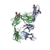
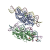
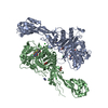
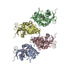
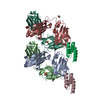
| Title | Structure of the FliH-FliI complex | ||||||
 Components Components |
| ||||||
 Keywords Keywords | HYDROLASE/MOTOR PROTEIN / Bacterial flagellum / type III secretion / ATPase / peripheral stalk / HYDROLASE-MOTOR PROTEIN complex | ||||||
| Function / homology |  Function and homology information Function and homology informationbacterial-type flagellum organization / type III protein secretion system complex / protein secretion by the type III secretion system / bacterial-type flagellum / proton motive force-driven ATP synthesis / cytoskeletal motor activity / bacterial-type flagellum assembly / bacterial-type flagellum-dependent cell motility / H+-transporting two-sector ATPase / proton-transporting ATP synthase complex ...bacterial-type flagellum organization / type III protein secretion system complex / protein secretion by the type III secretion system / bacterial-type flagellum / proton motive force-driven ATP synthesis / cytoskeletal motor activity / bacterial-type flagellum assembly / bacterial-type flagellum-dependent cell motility / H+-transporting two-sector ATPase / proton-transporting ATP synthase complex / proton transmembrane transport / protein transport / ATP hydrolysis activity / ATP binding / identical protein binding / cytoplasm / cytosol Similarity search - Function | ||||||
| Biological species |  Salmonella typhimurium (bacteria) Salmonella typhimurium (bacteria) | ||||||
| Method |  X-RAY DIFFRACTION / SYNCHROTRON / MOLECULAR REPLACEMENT / Resolution: 3 Å X-RAY DIFFRACTION / SYNCHROTRON / MOLECULAR REPLACEMENT / Resolution: 3 Å | ||||||
 Authors Authors | Imada, K. / Uchida, Y. / Kinoshita, M. / Namba, K. / Minamino, T. | ||||||
 Citation Citation | Journal: Proc.Natl.Acad.Sci.USA / Year: 2016 Title: Insight into the flagella type III export revealed by the complex structure of the type III ATPase and its regulator Authors: Imada, K. / Minamino, T. / Uchida, Y. / Kinoshita, M. / Namba, K. #1: Journal: Acta Crystallogr. Sect. F Struct. Biol. Cryst. Commun. Year: 2012 Title: Crystallization and preliminary X-ray analysis of the FliH-FliI complex responsible for bacterial flagellar type III protein export. Authors: Uchida, Y. / Minamino, T. / Namba, K. / Imada, K. | ||||||
| History |
|
- Structure visualization
| Structure viewer | Molecule: MolmilJmol/JSmol |
|---|
- Downloads & links
-Download
| PDBx/mmCIF format | 5b0o.cif.gz | 542.1 KB | Display | PDBx/mmCIF format |
|---|---|---|---|---|
| PDB format | pdb5b0o.ent.gz | 446.8 KB | Display | PDB format |
| PDBx/mmJSON format | 5b0o.json.gz | Tree view | PDBx/mmJSON format | |
| Others |  Other downloads Other downloads |
-Validation report
| Arichive directory | https://data.pdbj.org/pub/pdb/validation_reports/b0/5b0oftp://data.pdbj.org/pub/pdb/validation_reports/b0/5b0o | HTTPS FTP |
|---|
-Related structure data
| Related structure data |  2dpyS S: Starting model for refinement |
|---|---|
| Similar structure data |


-Links
 PDBj
PDBj


- Assembly
| Deposited unit | 
| ||||||||
|---|---|---|---|---|---|---|---|---|---|
| 1 | 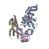
| ||||||||
| 2 | 
| ||||||||
| 3 | 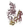
| ||||||||
| 4 | 
| ||||||||
| Unit cell |
|
-Components
| #1: Protein | Mass: 49319.551 Da / Num. of mol.: 4 Source method: isolated from a genetically manipulated source Source: (gene. exp.) Salmonella typhimurium (strain LT2 / SGSC1412 / ATCC 700720) (bacteria)Strain: LT2 / SGSC1412 / ATCC 700720 / Gene: fliI, fla AIII, flaC, STM1972 / Plasmid: pET15b / Production host: Escherichia coli / Strain (production host): BL21(DE3) References: UniProt: P26465, H+-transporting two-sector ATPase #2: Protein | Mass: 15335.442 Da / Num. of mol.: 8 / Fragment: UNP residues 99-235 Source method: isolated from a genetically manipulated source Source: (gene. exp.) Salmonella typhimurium (bacteria) / Strain: LT2 / SGSC1412 / ATCC 700720 / Gene: fliH, fla AII.3, fla BIII, STM1971 / Production host: #3: Chemical | ChemComp-ADP /   Mass: 427.201 Da / Num. of mol.: 4 / Source method: obtained synthetically / Formula: C10H15N5O10P2 / Comment: ADP, energy-carrying molecule*YM Mass: 427.201 Da / Num. of mol.: 4 / Source method: obtained synthetically / Formula: C10H15N5O10P2 / Comment: ADP, energy-carrying molecule*YM |
|---|
-Experimental details
-Experiment
| Experiment | Method: X-RAY DIFFRACTION |
|---|
- Sample preparation
| Crystal | Density Matthews: 2.57 Å3/Da / Density % sol: 52.13 % |
|---|---|
| Crystal grow | Temperature: 285 K / Method: vapor diffusion, sitting drop / pH: 7.2 Details: 5%(w/v) PEG400, 0.1M HEPES pH7.2 and 100mM Magnesium acetate |
-Data collection
| Diffraction | Mean temperature: 40 K / Ambient temp details: helium gas flow |
|---|---|
| Diffraction source | Source: SYNCHROTRON / Site: SPring-8  / Beamline: BL41XU / Wavelength: 1 Å / Beamline: BL41XU / Wavelength: 1 Å |
| Detector | Type: ADSC QUANTUM 315 / Detector: CCD / Date: Feb 3, 2010 |
| Radiation | Monochromator: Double-crystal monochromator / Protocol: SINGLE WAVELENGTH / Monochromatic (M) / Laue (L): M / Scattering type: x-ray |
| Radiation wavelength | Wavelength: 1 Å / Relative weight: 1 |
| Reflection | Resolution: 3→60 Å / Num. obs: 65054 / % possible obs: 99.4 % / Redundancy: 4.6 % / Rmerge(I) obs: 0.109 / Net I/σ(I): 9.4 |
| Reflection shell | Resolution: 3→3.16 Å / Redundancy: 4.2 % / Rmerge(I) obs: 0.362 / Mean I/σ(I) obs: 3.5 / % possible all: 99.6 |
- Processing
| Software |
| |||||||||||||||||||||||||||||||||||||||||||||||||||||||||||||||||||||||||||||||||||||||||||||||||||||||||||||||||||||||||||||||||||||||||||||||||||||||||||||||||||||||||||||||
|---|---|---|---|---|---|---|---|---|---|---|---|---|---|---|---|---|---|---|---|---|---|---|---|---|---|---|---|---|---|---|---|---|---|---|---|---|---|---|---|---|---|---|---|---|---|---|---|---|---|---|---|---|---|---|---|---|---|---|---|---|---|---|---|---|---|---|---|---|---|---|---|---|---|---|---|---|---|---|---|---|---|---|---|---|---|---|---|---|---|---|---|---|---|---|---|---|---|---|---|---|---|---|---|---|---|---|---|---|---|---|---|---|---|---|---|---|---|---|---|---|---|---|---|---|---|---|---|---|---|---|---|---|---|---|---|---|---|---|---|---|---|---|---|---|---|---|---|---|---|---|---|---|---|---|---|---|---|---|---|---|---|---|---|---|---|---|---|---|---|---|---|---|---|---|---|---|
| Refinement | Method to determine structure: MOLECULAR REPLACEMENT Starting model: 2DPY Resolution: 3→51.315 Å / SU ML: 0.96 / Cross valid method: THROUGHOUT / σ(F): 1.36 / Phase error: 25.8 / Stereochemistry target values: ML
| |||||||||||||||||||||||||||||||||||||||||||||||||||||||||||||||||||||||||||||||||||||||||||||||||||||||||||||||||||||||||||||||||||||||||||||||||||||||||||||||||||||||||||||||
| Solvent computation | Shrinkage radii: 1.06 Å / VDW probe radii: 1.3 Å / Solvent model: FLAT BULK SOLVENT MODEL / Bsol: 37.271 Å2 / ksol: 0.267 e/Å3 | |||||||||||||||||||||||||||||||||||||||||||||||||||||||||||||||||||||||||||||||||||||||||||||||||||||||||||||||||||||||||||||||||||||||||||||||||||||||||||||||||||||||||||||||
| Displacement parameters |
| |||||||||||||||||||||||||||||||||||||||||||||||||||||||||||||||||||||||||||||||||||||||||||||||||||||||||||||||||||||||||||||||||||||||||||||||||||||||||||||||||||||||||||||||
| Refinement step | Cycle: LAST / Resolution: 3→51.315 Å
| |||||||||||||||||||||||||||||||||||||||||||||||||||||||||||||||||||||||||||||||||||||||||||||||||||||||||||||||||||||||||||||||||||||||||||||||||||||||||||||||||||||||||||||||
| Refine LS restraints |
| |||||||||||||||||||||||||||||||||||||||||||||||||||||||||||||||||||||||||||||||||||||||||||||||||||||||||||||||||||||||||||||||||||||||||||||||||||||||||||||||||||||||||||||||
| LS refinement shell |
|
