 Movie
Movie Controller

[English] 日本語
 Yorodumi
- PDB-4ytz: Rat xanthine oxidoreductase, C-terminal deletion protein variant,... -
+ Open data

- Basic information
| Entry | Database: PDB / ID: 4ytz | ||||||
|---|---|---|---|---|---|---|---|
| Title | Rat xanthine oxidoreductase, C-terminal deletion protein variant, crystal grown without dithiothreitol | ||||||
 Components Components | Xanthine dehydrogenase/oxidase | ||||||
 Keywords Keywords | OXIDOREDUCTASE / xanthine oxidoreductase / xanthine oxidase / xanthine dehydrogenase / D/O conversion | ||||||
| Function / homology |  Function and homology information Function and homology informationButyrophilin (BTN) family interactions / hypoxanthine catabolic process / hypoxanthine dehydrogenase activity / hypoxanthine oxidase activity / Azathioprine ADME / Purine catabolism / guanine catabolic process / deoxyguanosine catabolic process / xanthine dehydrogenase / xanthine oxidase ...Butyrophilin (BTN) family interactions / hypoxanthine catabolic process / hypoxanthine dehydrogenase activity / hypoxanthine oxidase activity / Azathioprine ADME / Purine catabolism / guanine catabolic process / deoxyguanosine catabolic process / xanthine dehydrogenase / xanthine oxidase / xanthine oxidase activity / xanthine catabolic process / xanthine dehydrogenase activity / GMP catabolic process / deoxyinosine catabolic process / deoxyadenosine catabolic process / dAMP catabolic process / inosine catabolic process / regulation of epithelial cell differentiation / response to azide / : / adenosine catabolic process / response to aluminum ion / dGMP catabolic process / AMP catabolic process / IMP catabolic process / allantoin metabolic process / response to carbon monoxide / molybdopterin cofactor binding / nitrite reductase (NO-forming) activity / iron-sulfur cluster assembly / cellular response to interleukin-1 / lactation / FAD binding / sarcoplasmic reticulum / response to hydrogen peroxide / 2 iron, 2 sulfur cluster binding / cellular response to tumor necrosis factor / peroxisome / flavin adenine dinucleotide binding / response to lipopolysaccharide / oxidoreductase activity / iron ion binding / protein homodimerization activity / extracellular space / identical protein binding / cytosol Similarity search - Function | ||||||
| Biological species |  | ||||||
| Method |  X-RAY DIFFRACTION / SYNCHROTRON / MOLECULAR REPLACEMENT / molecular replacement / Resolution: 2.3 Å X-RAY DIFFRACTION / SYNCHROTRON / MOLECULAR REPLACEMENT / molecular replacement / Resolution: 2.3 Å | ||||||
 Authors Authors | Nishino, T. / Okamoto, K. / Kawaguchi, Y. / Matsumura, T. / Eger, B.T. / Pai, E.F. / Nishino, T. | ||||||
 Citation Citation | Journal: Febs J. / Year: 2015 Title: The C-terminal peptide plays a role in the formation of an intermediate form during the transition between xanthine dehydrogenase and xanthine oxidase Authors: Nishino, T. / Okamoto, K. / Kawaguchi, Y. / Matsumura, T. / Eger, B.T. / Pai, E.F. / Nishino, T. | ||||||
| History |
|
- Structure visualization
| Structure viewer | Molecule: MolmilJmol/JSmol |
|---|
- Downloads & links
-Download
| PDBx/mmCIF format | 4ytz.cif.gz | 506 KB | Display | PDBx/mmCIF format |
|---|---|---|---|---|
| PDB format | pdb4ytz.ent.gz | 404.6 KB | Display | PDB format |
| PDBx/mmJSON format | 4ytz.json.gz | Tree view | PDBx/mmJSON format | |
| Others |  Other downloads Other downloads |
-Validation report
| Arichive directory | https://data.pdbj.org/pub/pdb/validation_reports/yt/4ytzftp://data.pdbj.org/pub/pdb/validation_reports/yt/4ytz | HTTPS FTP |
|---|
-Related structure data
| Related structure data |  4yrwC  4yswC  4ytyC  3an1S C: citing same article ( S: Starting model for refinement |
|---|---|
| Similar structure data |


-Links
 PDBj
PDBj


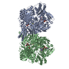
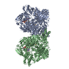
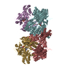
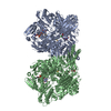
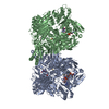
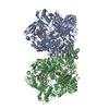
- Assembly
| Deposited unit | 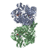
| ||||||||
|---|---|---|---|---|---|---|---|---|---|
| 1 |
| ||||||||
| Unit cell |
|
-Components
-Protein , 1 types, 2 molecules AB
| #1: Protein | Mass: 144652.344 Da / Num. of mol.: 2 / Fragment: UNP residues 1-1315 Source method: isolated from a genetically manipulated source Source: (gene. exp.)   Spodoptera frugiperda (fall armyworm) Spodoptera frugiperda (fall armyworm)References: UniProt: P22985, xanthine dehydrogenase, xanthine oxidase |
|---|
-Non-polymers , 6 types, 119 molecules 


| #2: Chemical | ChemComp-FES /  Mass: 175.820 Da / Num. of mol.: 4 / Source method: obtained synthetically / Formula: Fe2S2 Mass: 175.820 Da / Num. of mol.: 4 / Source method: obtained synthetically / Formula: Fe2S2#3: Chemical |  Mass: 785.550 Da / Num. of mol.: 2 / Source method: obtained synthetically / Formula: C27H33N9O15P2 / Comment: FAD*YM Mass: 785.550 Da / Num. of mol.: 2 / Source method: obtained synthetically / Formula: C27H33N9O15P2 / Comment: FAD*YM#4: Chemical |  Mass: 168.110 Da / Num. of mol.: 2 / Source method: obtained synthetically / Formula: C5H4N4O3 Mass: 168.110 Da / Num. of mol.: 2 / Source method: obtained synthetically / Formula: C5H4N4O3#5: Chemical |  Mass: 61.017 Da / Num. of mol.: 2 / Source method: obtained synthetically / Formula: CHO3 / Comment: pH buffer*YM Mass: 61.017 Da / Num. of mol.: 2 / Source method: obtained synthetically / Formula: CHO3 / Comment: pH buffer*YM#6: Chemical | ChemComp-CA /  Mass: 40.078 Da / Num. of mol.: 4 / Source method: obtained synthetically / Formula: Ca Mass: 40.078 Da / Num. of mol.: 4 / Source method: obtained synthetically / Formula: Ca#7: Water | ChemComp-HOH / | Mass: 18.015 Da / Num. of mol.: 105 / Source method: isolated from a natural source / Formula: H2O |
|---|
-Details
| Has protein modification | Y |
|---|
-Experimental details
-Experiment
| Experiment | Method: X-RAY DIFFRACTION / Number of used crystals: 1 |
|---|
- Sample preparation
| Crystal | Density Matthews: 2.75 Å3/Da / Density % sol: 55.32 % |
|---|---|
| Crystal grow | Temperature: 293 K / Method: vapor diffusion, hanging drop / pH: 6.2 Details: PEGl 8000, lithium formate, sodium salicylate, EDTA, glycerol, HEPES |
-Data collection
| Diffraction | Mean temperature: 100 K | ||||||||||||||||||||||||||||||||||||||||||||||||||||||||||||||||||
|---|---|---|---|---|---|---|---|---|---|---|---|---|---|---|---|---|---|---|---|---|---|---|---|---|---|---|---|---|---|---|---|---|---|---|---|---|---|---|---|---|---|---|---|---|---|---|---|---|---|---|---|---|---|---|---|---|---|---|---|---|---|---|---|---|---|---|---|
| Diffraction source | Source: SYNCHROTRON / Site: Photon Factory  / Beamline: AR-NW12A / Wavelength: 1 Å / Beamline: AR-NW12A / Wavelength: 1 Å | ||||||||||||||||||||||||||||||||||||||||||||||||||||||||||||||||||
| Detector | Type: ADSC QUANTUM 210r / Detector: CCD / Date: Dec 13, 2006 | ||||||||||||||||||||||||||||||||||||||||||||||||||||||||||||||||||
| Radiation | Protocol: SINGLE WAVELENGTH / Monochromatic (M) / Laue (L): M / Scattering type: x-ray | ||||||||||||||||||||||||||||||||||||||||||||||||||||||||||||||||||
| Radiation wavelength | Wavelength: 1 Å / Relative weight: 1 | ||||||||||||||||||||||||||||||||||||||||||||||||||||||||||||||||||
| Reflection | Resolution: 2.3→30 Å / Num. obs: 135086 / % possible obs: 97 % / Redundancy: 3.5 % / Rmerge(I) obs: 0.082 / Χ2: 1.011 / Net I/av σ(I): 15.242 / Net I/σ(I): 14.7 / Num. measured all: 468751 | ||||||||||||||||||||||||||||||||||||||||||||||||||||||||||||||||||
| Reflection shell | Diffraction-ID: 1 / Rejects: _
|
-Phasing
| Phasing | Method: molecular replacement |
|---|
- Processing
| Software |
| |||||||||||||||||||||||||||||||||||||||||||||||||||||||||||||||||
|---|---|---|---|---|---|---|---|---|---|---|---|---|---|---|---|---|---|---|---|---|---|---|---|---|---|---|---|---|---|---|---|---|---|---|---|---|---|---|---|---|---|---|---|---|---|---|---|---|---|---|---|---|---|---|---|---|---|---|---|---|---|---|---|---|---|---|
| Refinement | Method to determine structure: MOLECULAR REPLACEMENT Starting model: 3AN1 Resolution: 2.3→29.87 Å / Cor.coef. Fo:Fc: 0.944 / Cor.coef. Fo:Fc free: 0.911 / SU B: 7.389 / SU ML: 0.179 / Cross valid method: THROUGHOUT / σ(F): 0 / ESU R: 0.312 / ESU R Free: 0.246 / Stereochemistry target values: MAXIMUM LIKELIHOOD Details: HYDROGENS HAVE BEEN ADDED IN THE RIDING POSITIONS U VALUES : REFINED INDIVIDUALLY
| |||||||||||||||||||||||||||||||||||||||||||||||||||||||||||||||||
| Solvent computation | Ion probe radii: 0.8 Å / Shrinkage radii: 0.8 Å / VDW probe radii: 1.4 Å / Solvent model: MASK | |||||||||||||||||||||||||||||||||||||||||||||||||||||||||||||||||
| Displacement parameters | Biso max: 100.46 Å2 / Biso mean: 35.084 Å2 / Biso min: 11.38 Å2
| |||||||||||||||||||||||||||||||||||||||||||||||||||||||||||||||||
| Refinement step | Cycle: final / Resolution: 2.3→29.87 Å
| |||||||||||||||||||||||||||||||||||||||||||||||||||||||||||||||||
| Refine LS restraints |
| |||||||||||||||||||||||||||||||||||||||||||||||||||||||||||||||||
| LS refinement shell | Resolution: 2.297→2.356 Å / Total num. of bins used: 20
|
