 Movie
Movie Controller

[English] 日本語
 Yorodumi
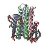
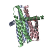
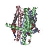
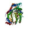
- PDB-4up6: Crystal structure of the wild-type diacylglycerol kinase refolded... -
+ Open data

- Basic information
| Entry | Database: PDB / ID: 4up6 | ||||||
|---|---|---|---|---|---|---|---|
| Title | Crystal structure of the wild-type diacylglycerol kinase refolded in the lipid cubic phase | ||||||
 Components Components | DIACYLGLYCEROL KINASE | ||||||
 Keywords Keywords | TRANSFERASE / 7.8 MAG / IN MESO / IN VITRO FOLDING / LIPID CUBIC PHASE / MEMBRANE PROTEIN / MONOACYLGLYCEROL / REFOLDING / RENATURATION | ||||||
| Function / homology |  Function and homology information Function and homology informationdiacylglycerol kinase (ATP) / lipid kinase activity / ATP-dependent diacylglycerol kinase activity / phosphatidic acid biosynthetic process / response to UV / ATP binding / metal ion binding / identical protein binding / membrane / plasma membrane Similarity search - Function | ||||||
| Biological species |  | ||||||
| Method |  X-RAY DIFFRACTION / SYNCHROTRON / MOLECULAR REPLACEMENT / Resolution: 3.801 Å X-RAY DIFFRACTION / SYNCHROTRON / MOLECULAR REPLACEMENT / Resolution: 3.801 Å | ||||||
 Authors Authors | Li, D. / Caffrey, M. | ||||||
 Citation Citation | Journal: Sci.Rep. / Year: 2014 Title: Renaturing Membrane Proteins in the Lipid Cubic Phase, a Nanoporous Membrane Mimetic. Authors: Li, D. / Caffrey, M. | ||||||
| History |
|
- Structure visualization
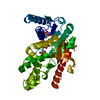
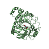
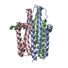
| Structure viewer | Molecule: MolmilJmol/JSmol |
|---|
- Downloads & links
-Download
| PDBx/mmCIF format | 4up6.cif.gz | 70.8 KB | Display | PDBx/mmCIF format |
|---|---|---|---|---|
| PDB format | pdb4up6.ent.gz | 54.2 KB | Display | PDB format |
| PDBx/mmJSON format | 4up6.json.gz | Tree view | PDBx/mmJSON format | |
| Others |  Other downloads Other downloads |
-Validation report
| Arichive directory | https://data.pdbj.org/pub/pdb/validation_reports/up/4up6ftp://data.pdbj.org/pub/pdb/validation_reports/up/4up6 | HTTPS FTP |
|---|
-Related structure data
| Related structure data |  4brbC  3ze4S S: Starting model for refinement C: citing same article ( |
|---|---|
| Similar structure data |


-Links
 PDBj
PDBj- Assembly
| Deposited unit | 
| ||||||||
|---|---|---|---|---|---|---|---|---|---|
| 1 |
| ||||||||
| Unit cell |
|
-Components
| #1: Protein | Mass: 14252.625 Da / Num. of mol.: 3 Source method: isolated from a genetically manipulated source Source: (gene. exp.) Sequence details | AN N-TERMINAL TAG (GHHHHHHEL) WAS ADDED TO AID PROTEIN PURIFICATION. THE N-TERMINAL MET IS CLEAVED, ...AN N-TERMINAL TAG (GHHHHHHEL) WAS ADDED TO AID PROTEIN PURIFICATI | |
|---|
-Experimental details
-Experiment
| Experiment | Method: X-RAY DIFFRACTION / Number of used crystals: 1 |
|---|
- Sample preparation
| Crystal | Density Matthews: 3.99 Å3/Da / Density % sol: 69.23 % / Description: NONE |
|---|---|
| Crystal grow | Temperature: 277 K / Method: lipidic cubic phase / pH: 5.6 Details: 3-5 %(V/V) 2-METHYL-2, 4-PENTANEDIOL, 0.1 M SODIUM CHLORIDE, 0.1 M LITHIUM NITRATE, 0.1 M SODIUM CITRATE/HCL PH 5.6. CRYSTALLIZED USING THE IN MESO (LIPIDIC CUBIC PHASE) METHOD AT 4 DEGREE ...Details: 3-5 %(V/V) 2-METHYL-2, 4-PENTANEDIOL, 0.1 M SODIUM CHLORIDE, 0.1 M LITHIUM NITRATE, 0.1 M SODIUM CITRATE/HCL PH 5.6. CRYSTALLIZED USING THE IN MESO (LIPIDIC CUBIC PHASE) METHOD AT 4 DEGREE CELSIUS WITH THE 7.8 MONOACYLGLYCEROL (7.8 MAG) AS THE HOSTING LIPID. |
-Data collection
| Diffraction | Mean temperature: 100 K |
|---|---|
| Diffraction source | Source: SYNCHROTRON / Site: APS  / Beamline: 23-ID-B / Wavelength: 1.0332 / Beamline: 23-ID-B / Wavelength: 1.0332 |
| Detector | Type: MARRESEARCH / Detector: CCD / Date: Feb 16, 2012 / Details: K-B PAIR OF BIOMORPH MIRRORS |
| Radiation | Monochromator: M / Protocol: SINGLE WAVELENGTH / Monochromatic (M) / Laue (L): M / Scattering type: x-ray |
| Radiation wavelength | Wavelength: 1.0332 Å / Relative weight: 1 |
| Reflection | Resolution: 3.8→54.44 Å / Num. obs: 6532 / % possible obs: 95.5 % / Observed criterion σ(I): -3 / Redundancy: 6.4 % / Biso Wilson estimate: 136.72 Å2 / Rmerge(I) obs: 0.14 / Net I/σ(I): 9.2 |
| Reflection shell | Resolution: 3.8→3.9 Å / Redundancy: 6.5 % / Rmerge(I) obs: 1.13 / Mean I/σ(I) obs: 1.7 / % possible all: 97.8 |
- Processing
| Software |
| ||||||||||||||||||||||||
|---|---|---|---|---|---|---|---|---|---|---|---|---|---|---|---|---|---|---|---|---|---|---|---|---|---|
| Refinement | Method to determine structure: MOLECULAR REPLACEMENT Starting model: PDB ENTRY 3ZE4 Resolution: 3.801→46.323 Å / SU ML: 0.59 / σ(F): 1.34 / Phase error: 43.17 / Stereochemistry target values: ML Details: THERE ARE THREE IDENTICAL MONOMERS IN THE ASYMMETRIC UNIT. NCS RESTRAINTS WERE APPLIED FOR TORSION ANGLES DURING REFINEMENT.
| ||||||||||||||||||||||||
| Solvent computation | Shrinkage radii: 0.9 Å / VDW probe radii: 1.11 Å / Solvent model: FLAT BULK SOLVENT MODEL | ||||||||||||||||||||||||
| Displacement parameters | Biso mean: 145.86 Å2 | ||||||||||||||||||||||||
| Refinement step | Cycle: LAST / Resolution: 3.801→46.323 Å
| ||||||||||||||||||||||||
| Refine LS restraints |
| ||||||||||||||||||||||||
| LS refinement shell |
|
