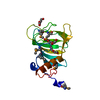
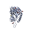
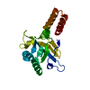
Entry Database : PDB / ID : 4uonTitle Crystal structure of C-terminal truncated (110-265) Aura virus capsid protease. CAPSID PROTEASE Keywords Function / homology Function Domain/homology Component
/ / / / / / / / / / / / / / / / / / / / / / / / / / / / / / / / / / / / / / / / / / / / / / / / / / / / / / / Biological species Method / / Resolution : 1.81 Å Authors Aggarwal, M. / Kumar, P. / Tomar, S. Journal : J.Virol. / Year : 2014Title : Trans-Protease Activity and Structural Insights Into the Active Form of the Alphavirus Capsid Protease.Authors : Aggarwal, M. / Dhindwal, S. / Kumar, P. / Kuhn, R.J. / Tomar, S. History Deposition Jun 5, 2014 Deposition site / Processing site Supersession Jun 18, 2014 ID 4AUS Revision 1.0 Jun 18, 2014 Provider / Type Revision 1.1 Oct 22, 2014 Group Revision 1.2 Jan 10, 2024 Group Data collection / Database references ... Data collection / Database references / Derived calculations / Other / Refinement description Category chem_comp_atom / chem_comp_bond ... chem_comp_atom / chem_comp_bond / database_2 / pdbx_database_status / pdbx_initial_refinement_model / struct_sheet / struct_site Item _database_2.pdbx_DOI / _database_2.pdbx_database_accession ... _database_2.pdbx_DOI / _database_2.pdbx_database_accession / _pdbx_database_status.status_code_sf / _struct_sheet.number_strands / _struct_site.pdbx_auth_asym_id / _struct_site.pdbx_auth_comp_id / _struct_site.pdbx_auth_seq_id
Show all Show less
 Movie
Movie Controller

 Yorodumi
 Open data
 Basic information
 Components
 Keywords
 Function and homology information
 AURA VIRUS
 X-RAY DIFFRACTION /
  Authors
 Citation
 Structure visualization
 Downloads & links
 Other downloads
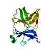


 PDBj
PDBj


 Assembly


 Mass: 92.094 Da / Num. of mol.: 6 / Source method: obtained synthetically / Formula: C3H8O3
 Mass: 18.015 Da / Num. of mol.: 400 / Source method: isolated from a natural source / Formula: H2O
 Sample preparation
 Processing