 Movie
Movie Controller

[English] 日本語
 Yorodumi
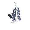
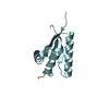
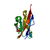
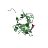
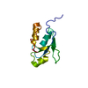
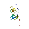
- PDB-4qy7: Crystal structure of a yobA protein (BSU18810) from Bacillus subt... -
+ Open data

- Basic information
| Entry | Database: PDB / ID: 4qy7 | ||||||
|---|---|---|---|---|---|---|---|
| Title | Crystal structure of a yobA protein (BSU18810) from Bacillus subtilis subsp. subtilis str. 168 at 1.55 A resolution | ||||||
 Components Components | Uncharacterized protein YobA | ||||||
 Keywords Keywords | STRUCTURAL GENOMICS / UNKNOWN FUNCTION / PF11518 family protein / DUF3221 / OB-fold / Joint Center for Structural Genomics / JCSG / Protein Structure Initiative / PSI-BIOLOGY | ||||||
| Function / homology | Protein of unknown function DUF3221 / Protein of unknown function (DUF3221) / Nucleic acid-binding proteins / OB fold (Dihydrolipoamide Acetyltransferase, E2P) / Nucleic acid-binding, OB-fold / Beta Barrel / Mainly Beta / Uncharacterized protein YobA Function and homology information Function and homology information | ||||||
| Biological species |  | ||||||
| Method |  X-RAY DIFFRACTION / SYNCHROTRON / MAD / Resolution: 1.55 Å X-RAY DIFFRACTION / SYNCHROTRON / MAD / Resolution: 1.55 Å | ||||||
 Authors Authors | Joint Center for Structural Genomics (JCSG) | ||||||
 Citation Citation | Journal: To be published Title: Crystal structure of a hypothetical protein (yobA) from Bacillus subtilis subsp. subtilis str. 168 at 1.55 A resolution Authors: Joint Center for Structural Genomics (JCSG) | ||||||
| History |
|
- Structure visualization
| Structure viewer | Molecule: MolmilJmol/JSmol |
|---|
- Downloads & links
-Download
| PDBx/mmCIF format | 4qy7.cif.gz | 52.5 KB | Display | PDBx/mmCIF format |
|---|---|---|---|---|
| PDB format | pdb4qy7.ent.gz | 39.3 KB | Display | PDB format |
| PDBx/mmJSON format | 4qy7.json.gz | Tree view | PDBx/mmJSON format | |
| Others |  Other downloads Other downloads |
-Validation report
| Summary document | 4qy7_validation.pdf.gz | 590.8 KB | Display | wwPDB validaton report |
|---|---|---|---|---|
| Full document | 4qy7_full_validation.pdf.gz | 590.8 KB | Display | |
| Data in XML | 4qy7_validation.xml.gz | 7 KB | Display | |
| Data in CIF | 4qy7_validation.cif.gz | 9.1 KB | Display | |
| Arichive directory | https://data.pdbj.org/pub/pdb/validation_reports/qy/4qy7ftp://data.pdbj.org/pub/pdb/validation_reports/qy/4qy7 | HTTPS FTP |
-Related structure data
| Similar structure data | |
|---|---|
| Other databases |
-Links
 PDBj
PDBj- Assembly
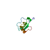
| Deposited unit | 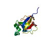
| ||||||||
|---|---|---|---|---|---|---|---|---|---|
| 1 |
| ||||||||
| Unit cell |
| ||||||||
| Details | CRYSTAL PACKING ANALYSIS SUGGESTS THE ASSIGNMENT OF A MONOMER AS THE SIGNIFICANT OLIGOMERIZATION STATE. |
-Components
| #1: Protein | Mass: 10690.822 Da / Num. of mol.: 1 Source method: isolated from a genetically manipulated source Source: (gene. exp.) Strain: 168 / Gene: yobA, BSU18810 / Plasmid: SpeedET / Production host: |
|---|---|
| #2: Chemical | ChemComp-2PE /   Mass: 414.488 Da / Num. of mol.: 1 / Source method: obtained synthetically / Formula: C18H38O10 / Comment: precipitant*YM Mass: 414.488 Da / Num. of mol.: 1 / Source method: obtained synthetically / Formula: C18H38O10 / Comment: precipitant*YM |
| #3: Water | ChemComp-HOH /  Mass: 18.015 Da / Num. of mol.: 89 / Source method: isolated from a natural source / Formula: H2O Mass: 18.015 Da / Num. of mol.: 89 / Source method: isolated from a natural source / Formula: H2O |
| Sequence details | THIS CONSTRUCT WAS EXPRESSED WITH AN N-TERMINAL PURIFICATION TAG MGSDKIHHHHHHENLYFQG. THE TAG WAS ...THIS CONSTRUCT WAS EXPRESSED WITH AN N-TERMINAL PURIFICATI |
-Experimental details
-Experiment
| Experiment | Method: X-RAY DIFFRACTION / Number of used crystals: 1 |
|---|
- Sample preparation
| Crystal | Density Matthews: 1.99 Å3/Da / Density % sol: 38.15 % |
|---|---|
| Crystal grow | Temperature: 277 K / Method: vapor diffusion, sitting drop / pH: 6 Details: 5.00% polyethylene glycol 3000, 40.00% polyethylene glycol 400, 0.1M MES pH 6.0, NANODROP, VAPOR DIFFUSION, SITTING DROP, temperature 277K |
-Data collection
| Diffraction | Mean temperature: 100 K | |||||||||||||||||||||||||||||||||||||||||||||||||||||||||||||||||||||||||||||
|---|---|---|---|---|---|---|---|---|---|---|---|---|---|---|---|---|---|---|---|---|---|---|---|---|---|---|---|---|---|---|---|---|---|---|---|---|---|---|---|---|---|---|---|---|---|---|---|---|---|---|---|---|---|---|---|---|---|---|---|---|---|---|---|---|---|---|---|---|---|---|---|---|---|---|---|---|---|---|
| Diffraction source | Source: SYNCHROTRON / Site: SSRL  / Beamline: BL14-1 / Wavelength: 0.95369,0.97937,0.97922 / Beamline: BL14-1 / Wavelength: 0.95369,0.97937,0.97922 | |||||||||||||||||||||||||||||||||||||||||||||||||||||||||||||||||||||||||||||
| Detector | Type: MARMOSAIC 325 mm CCD / Detector: CCD / Date: Jun 11, 2014 Details: Vertical focusing mirror; double crystal Si(111) monochromator | |||||||||||||||||||||||||||||||||||||||||||||||||||||||||||||||||||||||||||||
| Radiation | Monochromator: double crystal Si(111) / Protocol: MAD / Monochromatic (M) / Laue (L): M / Scattering type: x-ray | |||||||||||||||||||||||||||||||||||||||||||||||||||||||||||||||||||||||||||||
| Radiation wavelength |
| |||||||||||||||||||||||||||||||||||||||||||||||||||||||||||||||||||||||||||||
| Reflection | Resolution: 1.55→27.746 Å / Num. obs: 12907 / % possible obs: 98.3 % / Observed criterion σ(I): -3 / Biso Wilson estimate: 21.025 Å2 / Rmerge(I) obs: 0.031 / Net I/σ(I): 14.83 | |||||||||||||||||||||||||||||||||||||||||||||||||||||||||||||||||||||||||||||
| Reflection shell |
|
-Phasing
| Phasing | Method: MAD |
|---|
- Processing
| Software |
| ||||||||||||||||||||||||||||||||||||||||||||||||||||||||||||||||||||||||||||||||||||||||||||||||||||||||||||
|---|---|---|---|---|---|---|---|---|---|---|---|---|---|---|---|---|---|---|---|---|---|---|---|---|---|---|---|---|---|---|---|---|---|---|---|---|---|---|---|---|---|---|---|---|---|---|---|---|---|---|---|---|---|---|---|---|---|---|---|---|---|---|---|---|---|---|---|---|---|---|---|---|---|---|---|---|---|---|---|---|---|---|---|---|---|---|---|---|---|---|---|---|---|---|---|---|---|---|---|---|---|---|---|---|---|---|---|---|---|
| Refinement | Method to determine structure: MAD / Resolution: 1.55→27.746 Å / Cor.coef. Fo:Fc: 0.9571 / Cor.coef. Fo:Fc free: 0.939 / Occupancy max: 1 / Occupancy min: 0.26 / Cross valid method: THROUGHOUT / σ(F): 0 Details: 1. ZERO OCCUPANCY HYDROGENS WERE INCLUDED DURING REFINEMENT TO IMPROVE THE ANTI-BUMPING RESTRAINTS. 2. ATOM RECORDS CONTAIN SUM OF TLS AND RESIDUAL B FACTORS. 3. ANISOU RECORDS CONTAIN SUM ...Details: 1. ZERO OCCUPANCY HYDROGENS WERE INCLUDED DURING REFINEMENT TO IMPROVE THE ANTI-BUMPING RESTRAINTS. 2. ATOM RECORDS CONTAIN SUM OF TLS AND RESIDUAL B FACTORS. 3. ANISOU RECORDS CONTAIN SUM OF TLS AND RESIDUAL U FACTORS. 4. A MET-INHIBITION PROTOCOL WAS USED FOR SELENOMETHIONINE INCORPORATION DURING PROTEIN EXPRESSION. THE OCCUPANCY OF THE SE ATOMS IN THE MSE RESIDUES WAS REDUCED TO 0.75 FOR THE REDUCED SCATTERING POWER DUE TO PARTIAL S-MET INCORPORATION. 5. MAD PHASE RESTRAINTS WERE USED DURING REFINEMENT. 6. NONAETHYLENE GLYCOL (2PE) MOLECULE FROM THE CRYSTALLIZATION SOLUTION ARE MODELED.
| ||||||||||||||||||||||||||||||||||||||||||||||||||||||||||||||||||||||||||||||||||||||||||||||||||||||||||||
| Displacement parameters | Biso max: 97.54 Å2 / Biso mean: 28.8288 Å2 / Biso min: 11.53 Å2
| ||||||||||||||||||||||||||||||||||||||||||||||||||||||||||||||||||||||||||||||||||||||||||||||||||||||||||||
| Refine analyze | Luzzati coordinate error obs: 0.232 Å | ||||||||||||||||||||||||||||||||||||||||||||||||||||||||||||||||||||||||||||||||||||||||||||||||||||||||||||
| Refinement step | Cycle: LAST / Resolution: 1.55→27.746 Å
| ||||||||||||||||||||||||||||||||||||||||||||||||||||||||||||||||||||||||||||||||||||||||||||||||||||||||||||
| Refine LS restraints |
| ||||||||||||||||||||||||||||||||||||||||||||||||||||||||||||||||||||||||||||||||||||||||||||||||||||||||||||
| LS refinement shell | Resolution: 1.55→1.7 Å / Total num. of bins used: 6
| ||||||||||||||||||||||||||||||||||||||||||||||||||||||||||||||||||||||||||||||||||||||||||||||||||||||||||||
| Refinement TLS params. | Method: refined / Origin x: 7.7967 Å / Origin y: 7.1928 Å / Origin z: 8.7526 Å
|
