 Movie
Movie Controller

[English] 日本語
 Yorodumi
- PDB-3g19: The structure of the Caulobacter crescentus clpS protease adaptor... -
+ Open data

- Basic information
| Entry | Database: PDB / ID: 3g19 | ||||||
|---|---|---|---|---|---|---|---|
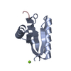
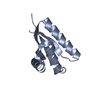
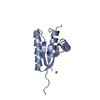
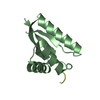
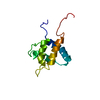
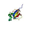
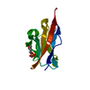
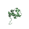
| Title | The structure of the Caulobacter crescentus clpS protease adaptor protein in complex with LLL tripeptide | ||||||
 Components Components |
| ||||||
 Keywords Keywords | PEPTIDE BINDING PROTEIN / adaptor / protein-peptide complex / peptide-binding protein | ||||||
| Function / homology |  Function and homology information Function and homology information | ||||||
| Biological species |  Caulobacter vibrioides (bacteria) Caulobacter vibrioides (bacteria) | ||||||
| Method |  X-RAY DIFFRACTION / MOLECULAR REPLACEMENT / molecular replacement / Resolution: 1.849 Å X-RAY DIFFRACTION / MOLECULAR REPLACEMENT / molecular replacement / Resolution: 1.849 Å | ||||||
 Authors Authors | Baker, T.A. / Roman-Hernandez, G. / Sauer, R.T. / Grant, R.A. | ||||||
 Citation Citation | Journal: Proc.Natl.Acad.Sci.USA / Year: 2009 Title: Molecular basis of substrate selection by the N-end rule adaptor protein ClpS. Authors: Roman-Hernandez, G. / Grant, R.A. / Sauer, R.T. / Baker, T.A. | ||||||
| History |
|
- Structure visualization
| Structure viewer | Molecule: MolmilJmol/JSmol |
|---|
- Downloads & links
-Download
| PDBx/mmCIF format | 3g19.cif.gz | 32.7 KB | Display | PDBx/mmCIF format |
|---|---|---|---|---|
| PDB format | pdb3g19.ent.gz | 21.2 KB | Display | PDB format |
| PDBx/mmJSON format | 3g19.json.gz | Tree view | PDBx/mmJSON format | |
| Others |  Other downloads Other downloads |
-Validation report
| Arichive directory | https://data.pdbj.org/pub/pdb/validation_reports/g1/3g19ftp://data.pdbj.org/pub/pdb/validation_reports/g1/3g19 | HTTPS FTP |
|---|
-Related structure data


-Links
 PDBj
PDBj

- Assembly
| Deposited unit | 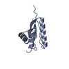
| ||||||||
|---|---|---|---|---|---|---|---|---|---|
| 1 |
| ||||||||
| Unit cell |
|
-Components
| #1: Protein | Mass: 9944.347 Da / Num. of mol.: 1 Source method: isolated from a genetically manipulated source Source: (gene. exp.) Caulobacter vibrioides (bacteria) / Strain: CB15 / Gene: CC_2467, clpS / Plasmid: pET23b / Production host: |
|---|---|
| #2: Protein/peptide | Mass: 357.489 Da / Num. of mol.: 1 / Source method: obtained synthetically / Details: synthetic peptide |
| #3: Water | ChemComp-HOH /  Mass: 18.015 Da / Num. of mol.: 127 / Source method: isolated from a natural source / Formula: H2O Mass: 18.015 Da / Num. of mol.: 127 / Source method: isolated from a natural source / Formula: H2O |
-Experimental details
-Experiment
| Experiment | Method: X-RAY DIFFRACTION / Number of used crystals: 1 |
|---|
- Sample preparation
| Crystal | Density Matthews: 1.71663 Å3/Da / Density % sol: 28.348003 % |
|---|---|
| Crystal grow | Temperature: 300 K / Method: vapor diffusion / pH: 5.5 Details: 0.1 M bis-tris pH 5.5, 0.2 M MgCl2, 25% PEG 3350, vapor diffusion, temperature 300K |
-Data collection
| Diffraction | Mean temperature: 100 K | ||||||||||||||||||||||||||||||||||||||||||||||||||||||||||||||||||
|---|---|---|---|---|---|---|---|---|---|---|---|---|---|---|---|---|---|---|---|---|---|---|---|---|---|---|---|---|---|---|---|---|---|---|---|---|---|---|---|---|---|---|---|---|---|---|---|---|---|---|---|---|---|---|---|---|---|---|---|---|---|---|---|---|---|---|---|
| Diffraction source | Source: ROTATING ANODE / Type: RIGAKU MICROMAX-007 HF / Wavelength: 1.5418 Å | ||||||||||||||||||||||||||||||||||||||||||||||||||||||||||||||||||
| Detector | Type: RIGAKU RAXIS IV / Detector: IMAGE PLATE / Date: Aug 10, 2008 / Details: Varimax-HR | ||||||||||||||||||||||||||||||||||||||||||||||||||||||||||||||||||
| Radiation | Monochromator: Varimax-HF / Protocol: SINGLE WAVELENGTH / Monochromatic (M) / Laue (L): M / Scattering type: x-ray | ||||||||||||||||||||||||||||||||||||||||||||||||||||||||||||||||||
| Radiation wavelength | Wavelength: 1.5418 Å / Relative weight: 1 | ||||||||||||||||||||||||||||||||||||||||||||||||||||||||||||||||||
| Reflection | Resolution: 1.85→50 Å / Num. obs: 5661 / % possible obs: 87.1 % / Redundancy: 6 % / Biso Wilson estimate: 13.78 Å2 / Rmerge(I) obs: 0.043 / Rrim(I) all: 0.043 / Χ2: 1.062 / Net I/av σ(I): 39.645 / Net I/σ(I): 16.2 / Num. measured all: 33923 | ||||||||||||||||||||||||||||||||||||||||||||||||||||||||||||||||||
| Reflection shell |
|
-Phasing
| Phasing | Method: molecular replacement |
|---|
- Processing
| Software |
| |||||||||||||||||||||||||||||||||||
|---|---|---|---|---|---|---|---|---|---|---|---|---|---|---|---|---|---|---|---|---|---|---|---|---|---|---|---|---|---|---|---|---|---|---|---|---|
| Refinement | Method to determine structure: MOLECULAR REPLACEMENT / Resolution: 1.849→22.388 Å / Occupancy max: 1 / Occupancy min: 0.52 / FOM work R set: 0.849 / SU ML: 0.22 / σ(F): 1.34 / Stereochemistry target values: ML
| |||||||||||||||||||||||||||||||||||
| Solvent computation | Shrinkage radii: 0.9 Å / VDW probe radii: 1.11 Å / Solvent model: FLAT BULK SOLVENT MODEL / Bsol: 44.543 Å2 / ksol: 0.38 e/Å3 | |||||||||||||||||||||||||||||||||||
| Displacement parameters | Biso max: 40.72 Å2 / Biso mean: 13.429 Å2 / Biso min: 0 Å2
| |||||||||||||||||||||||||||||||||||
| Refinement step | Cycle: LAST / Resolution: 1.849→22.388 Å
| |||||||||||||||||||||||||||||||||||
| Refine LS restraints |
| |||||||||||||||||||||||||||||||||||
| LS refinement shell | Refine-ID: X-RAY DIFFRACTION / Total num. of bins used: 4
|
