 Movie
Movie Controller


+ Open data

- Basic information
| Entry | Database: PDB / ID: 4pzg | ||||||
|---|---|---|---|---|---|---|---|
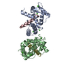
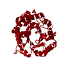
| Title | Crystal structure of human sorting nexin 10 (SNX10) | ||||||
 Components Components | Sorting nexin-10 | ||||||
 Keywords Keywords | PROTEIN TRANSPORT / sorting nexin / phox-homology domain | ||||||
| Function / homology |  Function and homology information Function and homology informationextrinsic component of endosome membrane / apical cytoplasm / tooth eruption / gastric acid secretion / 1-phosphatidylinositol binding / phosphatidylinositol phosphate binding / bone mineralization involved in bone maturation / vesicle organization / ruffle assembly / endosome organization ...extrinsic component of endosome membrane / apical cytoplasm / tooth eruption / gastric acid secretion / 1-phosphatidylinositol binding / phosphatidylinositol phosphate binding / bone mineralization involved in bone maturation / vesicle organization / ruffle assembly / endosome organization / cellular homeostasis / protein localization to cilium / protein localization to centrosome / cilium assembly / bone resorption / calcium ion homeostasis / osteoclast differentiation / secretory granule / cellular response to leukemia inhibitory factor / intracellular protein transport / endocytosis / ATPase binding / endosome / centrosome / endoplasmic reticulum / nucleus Similarity search - Function | ||||||
| Biological species |  Homo sapiens (human) Homo sapiens (human) | ||||||
| Method |  X-RAY DIFFRACTION / SYNCHROTRON / MOLECULAR REPLACEMENT / Resolution: 2.8 Å X-RAY DIFFRACTION / SYNCHROTRON / MOLECULAR REPLACEMENT / Resolution: 2.8 Å | ||||||
 Authors Authors | Xu, T. / Xu, J. / Wang, Q. / Liu, J. | ||||||
 Citation Citation | Journal: Proteins / Year: 2014 Title: Structure of human SNX10 reveals insights into its role in human autosomal recessive osteopetrosis. Authors: Xu, T. / Xu, J. / Ye, Y. / Wang, Q. / Shu, X. / Pei, D. / Liu, J. | ||||||
| History |
|
- Structure visualization
| Structure viewer | Molecule: MolmilJmol/JSmol |
|---|
- Downloads & links
-Download
| PDBx/mmCIF format | 4pzg.cif.gz | 78 KB | Display | PDBx/mmCIF format |
|---|---|---|---|---|
| PDB format | pdb4pzg.ent.gz | 62.2 KB | Display | PDB format |
| PDBx/mmJSON format | 4pzg.json.gz | Tree view | PDBx/mmJSON format | |
| Others |  Other downloads Other downloads |
-Validation report
| Summary document | 4pzg_validation.pdf.gz | 1.8 MB | Display | wwPDB validaton report |
|---|---|---|---|---|
| Full document | 4pzg_full_validation.pdf.gz | 1.8 MB | Display | |
| Data in XML | 4pzg_validation.xml.gz | 14.7 KB | Display | |
| Data in CIF | 4pzg_validation.cif.gz | 19 KB | Display | |
| Arichive directory | https://data.pdbj.org/pub/pdb/validation_reports/pz/4pzgftp://data.pdbj.org/pub/pdb/validation_reports/pz/4pzg | HTTPS FTP |
-Related structure data


-Links
 PDBj
PDBj- Assembly
| Deposited unit | 
| |||||||||||||||||||||||||||
|---|---|---|---|---|---|---|---|---|---|---|---|---|---|---|---|---|---|---|---|---|---|---|---|---|---|---|---|---|
| 1 |
| |||||||||||||||||||||||||||
| Unit cell |
| |||||||||||||||||||||||||||
| Components on special symmetry positions |
| |||||||||||||||||||||||||||
| Noncrystallographic symmetry (NCS) | NCS domain:
NCS domain segments:
|
-Components
| #1: Protein | Mass: 24701.484 Da / Num. of mol.: 2 Source method: isolated from a genetically manipulated source Source: (gene. exp.) Homo sapiens (human) / Gene: SNX10 / Production host:  #2: Chemical | ChemComp-NO3 /   Mass: 62.005 Da / Num. of mol.: 11 / Source method: obtained synthetically / Formula: NO3 Mass: 62.005 Da / Num. of mol.: 11 / Source method: obtained synthetically / Formula: NO3#3: Chemical | ChemComp-PE5 /   Mass: 398.489 Da / Num. of mol.: 7 / Source method: obtained synthetically / Formula: C18H38O9 / Comment: precipitant*YM Mass: 398.489 Da / Num. of mol.: 7 / Source method: obtained synthetically / Formula: C18H38O9 / Comment: precipitant*YM#4: Water | ChemComp-HOH / |  Mass: 18.015 Da / Num. of mol.: 15 / Source method: isolated from a natural source / Formula: H2O Mass: 18.015 Da / Num. of mol.: 15 / Source method: isolated from a natural source / Formula: H2O |
|---|
-Experimental details
-Experiment
| Experiment | Method: X-RAY DIFFRACTION / Number of used crystals: 1 |
|---|
- Sample preparation
| Crystal | Density Matthews: 5.24 Å3/Da / Density % sol: 76.53 % |
|---|---|
| Crystal grow | Temperature: 293 K / Method: vapor diffusion / pH: 7 Details: 4 M sodium nitrate, 0.1 M BIS-TRIS propane pH 7.0, vapor diffusion, temperature 293K |
-Data collection
| Diffraction source | Source: SYNCHROTRON / Site: SSRF  / Beamline: BL17U / Wavelength: 0.9792 Å / Beamline: BL17U / Wavelength: 0.9792 Å | |||||||||||||||||||||
|---|---|---|---|---|---|---|---|---|---|---|---|---|---|---|---|---|---|---|---|---|---|---|
| Detector | Type: ADSC QUANTUM 315 / Detector: CCD / Date: Mar 13, 2014 | |||||||||||||||||||||
| Radiation | Protocol: SINGLE WAVELENGTH / Monochromatic (M) / Laue (L): M / Scattering type: x-ray | |||||||||||||||||||||
| Radiation wavelength | Wavelength: 0.9792 Å / Relative weight: 1 | |||||||||||||||||||||
| Reflection | Resolution: 2.8→49.45 Å / Num. obs: 25144 / % possible obs: 98.7 % / Redundancy: 6.5 % / Rmerge(I) obs: 0.136 / Net I/σ(I): 10.5 / Scaling rejects: 121 | |||||||||||||||||||||
| Reflection shell | Diffraction-ID: 1
|
- Processing
| Software |
| ||||||||||||||||||||||||||||||||||||||||||||||||||||||||||||
|---|---|---|---|---|---|---|---|---|---|---|---|---|---|---|---|---|---|---|---|---|---|---|---|---|---|---|---|---|---|---|---|---|---|---|---|---|---|---|---|---|---|---|---|---|---|---|---|---|---|---|---|---|---|---|---|---|---|---|---|---|---|
| Refinement | Method to determine structure: MOLECULAR REPLACEMENT / Resolution: 2.8→49.45 Å / Cor.coef. Fo:Fc: 0.929 / Cor.coef. Fo:Fc free: 0.894 / SU B: 8.622 / SU ML: 0.164 / Cross valid method: THROUGHOUT / σ(F): 0 / ESU R: 0.25 / ESU R Free: 0.227 / Stereochemistry target values: MAXIMUM LIKELIHOOD Details: HYDROGENS HAVE BEEN ADDED IN THE RIDING POSITIONS U VALUES: REFINED INDIVIDUALLY
| ||||||||||||||||||||||||||||||||||||||||||||||||||||||||||||
| Solvent computation | Ion probe radii: 0.8 Å / Shrinkage radii: 0.8 Å / VDW probe radii: 1.2 Å / Solvent model: MASK | ||||||||||||||||||||||||||||||||||||||||||||||||||||||||||||
| Displacement parameters | Biso max: 152.81 Å2 / Biso mean: 42.045 Å2 / Biso min: 10.03 Å2
| ||||||||||||||||||||||||||||||||||||||||||||||||||||||||||||
| Refinement step | Cycle: LAST / Resolution: 2.8→49.45 Å
| ||||||||||||||||||||||||||||||||||||||||||||||||||||||||||||
| Refine LS restraints |
| ||||||||||||||||||||||||||||||||||||||||||||||||||||||||||||
| Refine LS restraints NCS | Ens-ID: 1 / Number: 8806 / Refine-ID: X-RAY DIFFRACTION / Type: interatomic distance / Rms dev position: 0.15 Å / Weight position: 0.05
| ||||||||||||||||||||||||||||||||||||||||||||||||||||||||||||
| LS refinement shell | Resolution: 2.8→2.873 Å / Total num. of bins used: 20
|
