 Movie
Movie Controller


+ Open data

- Basic information
| Entry | Database: PDB / ID: 4mon | ||||||
|---|---|---|---|---|---|---|---|
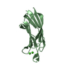
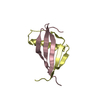
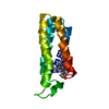
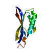
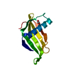
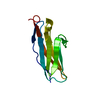
| Title | ORTHORHOMBIC MONELLIN | ||||||
 Components Components | (MONELLIN) x 2 | ||||||
 Keywords Keywords | SWEET-TASTING PROTEIN / ORTHORHOMBIC CRYSTALS | ||||||
| Function / homology |  Function and homology information Function and homology informationN-terminal domain of TfIIb - #130 / Helix Hairpins - #2000 / Monellin, A chain / Monellin, A chain superfamily / Monellin, B chain / : / Monellin / Monellin / N-terminal domain of TfIIb / Cystatin superfamily ...N-terminal domain of TfIIb - #130 / Helix Hairpins - #2000 / Monellin, A chain / Monellin, A chain superfamily / Monellin, B chain / : / Monellin / Monellin / N-terminal domain of TfIIb / Cystatin superfamily / Other non-globular / Helix Hairpins / Helix non-globular / Special Similarity search - Domain/homology | ||||||
| Biological species |  Dioscoreophyllum cumminsii (serendipity berry) Dioscoreophyllum cumminsii (serendipity berry) | ||||||
| Method |  X-RAY DIFFRACTION / MOLECULAR REPLACEMENT / Resolution: 2.3 Å X-RAY DIFFRACTION / MOLECULAR REPLACEMENT / Resolution: 2.3 Å | ||||||
 Authors Authors | Bujacz, G. / Wlodawer, A. | ||||||
 Citation Citation | Journal: Acta Crystallogr.,Sect.D / Year: 1997 Title: Structure of monellin refined to 2.3 a resolution in the orthorhombic crystal form. Authors: Bujacz, G. / Miller, M. / Harrison, R. / Thanki, N. / Gilliland, G.L. / Ogata, C.M. / Kim, S.H. / Wlodawer, A. | ||||||
| History |
|
- Structure visualization
| Structure viewer | Molecule: MolmilJmol/JSmol |
|---|
- Downloads & links
-Download
| PDBx/mmCIF format | 4mon.cif.gz | 54.7 KB | Display | PDBx/mmCIF format |
|---|---|---|---|---|
| PDB format | pdb4mon.ent.gz | 41.2 KB | Display | PDB format |
| PDBx/mmJSON format | 4mon.json.gz | Tree view | PDBx/mmJSON format | |
| Others |  Other downloads Other downloads |
-Validation report
| Arichive directory | https://data.pdbj.org/pub/pdb/validation_reports/mo/4monftp://data.pdbj.org/pub/pdb/validation_reports/mo/4mon | HTTPS FTP |
|---|
-Related structure data
| Related structure data |  3monS S: Starting model for refinement |
|---|---|
| Similar structure data |


-Links
 PDBj
PDBj
- Assembly
| Deposited unit | 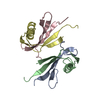
| ||||||||
|---|---|---|---|---|---|---|---|---|---|
| 1 | 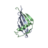
| ||||||||
| 2 | 
| ||||||||
| Unit cell |
|
-Components
| #1: Protein/peptide | Mass: 5407.167 Da / Num. of mol.: 2 / Source method: isolated from a natural source / Details: PROTEIN EXTRACTED FROM BERRIES Source: (natural) Dioscoreophyllum cumminsii (serendipity berry)References: UniProt: P02881 #2: Protein/peptide | Mass: 5841.647 Da / Num. of mol.: 2 / Source method: isolated from a natural source / Details: PROTEIN EXTRACTED FROM BERRIES Source: (natural) Dioscoreophyllum cumminsii (serendipity berry)References: UniProt: P02882 #3: Water | ChemComp-HOH / |  Mass: 18.015 Da / Num. of mol.: 180 / Source method: isolated from a natural source / Formula: H2O Mass: 18.015 Da / Num. of mol.: 180 / Source method: isolated from a natural source / Formula: H2OSequence details | PHE 1, RESIDUE PHE 1, NOTED IN SWISS-PROT P02881 AS {VARIANT...MISSING (IN 90% OF THE CHAINS)}, IS ...PHE 1, RESIDUE PHE 1, NOTED IN SWISS-PROT P02881 AS {VARIANT...MISSING (IN 90% OF THE CHAINS)}, IS NOT VISIBLE IN THIS STRUCTURE. THE SEQUENCE OF THE LAST TWO RESIDUES OF THE B/D CHAIN HAS BEEN REMARK 999 GIVEN AS EITHER ASNGLU OR GLUASN, DEPENDING ON THE SOURCE OF THE DATA. THE SEQUENCE USED HERE WAS PREVIOUSLY | |
|---|
-Experimental details
-Experiment
| Experiment | Method: X-RAY DIFFRACTION / Number of used crystals: 1 |
|---|
- Sample preparation
| Crystal | Density Matthews: 2.84 Å3/Da / Density % sol: 54.45 % Description: REJECTION CRITERIA DEFAULT FOR DATA COLLECTION USING XENGEN IS -3 SIGMA; THIS VALUE WAS NOT ALLOWED BY PDB DEPOSITION SOFTWARE. | ||||||||||||||||||||||||
|---|---|---|---|---|---|---|---|---|---|---|---|---|---|---|---|---|---|---|---|---|---|---|---|---|---|
| Crystal grow | pH: 7.2 Details: 5-10 MG/ML PROTEIN DISSOLVED IN 100 MM SODIUM PHOSPHATE BUFFER, PH 7.2, DIFFUSED AGAINST 20 W/V % ETHANOL. CRYSTALS UNSTABLE- CROSSLINKED WITH 0.015% GLUTARALDEHYDE FOR 1-1.5 HOURS. | ||||||||||||||||||||||||
| Crystal grow | *PLUS Method: vapor diffusion, hanging drop | ||||||||||||||||||||||||
| Components of the solutions | *PLUS
|
-Data collection
| Diffraction | Mean temperature: 290 K |
|---|---|
| Diffraction source | Source: ROTATING ANODE / Type: ELLIOTT GX-21 / Wavelength: 1.5418 |
| Detector | Type: SIEMENS / Detector: AREA DETECTOR / Date: Sep 1, 1985 / Details: COLLIMATOR |
| Radiation | Monochromator: GRAPHITE(002) / Monochromatic (M) / Laue (L): M / Scattering type: x-ray |
| Radiation wavelength | Wavelength: 1.5418 Å / Relative weight: 1 |
| Reflection | Resolution: 2.3→99 Å / Num. obs: 10602 / % possible obs: 90.9 % / Observed criterion σ(I): 0 / Redundancy: 3.4 % / Rsym value: 0.066 |
| Reflection | *PLUS Num. all: 11667 / Num. measured all: 36281 / Rmerge(I) obs: 0.066 |
- Processing
| Software |
| ||||||||||||||||||||||||||||||||||||||||||||||||||||||||||||||||||||||||||||||||||||
|---|---|---|---|---|---|---|---|---|---|---|---|---|---|---|---|---|---|---|---|---|---|---|---|---|---|---|---|---|---|---|---|---|---|---|---|---|---|---|---|---|---|---|---|---|---|---|---|---|---|---|---|---|---|---|---|---|---|---|---|---|---|---|---|---|---|---|---|---|---|---|---|---|---|---|---|---|---|---|---|---|---|---|---|---|---|
| Refinement | Method to determine structure: MOLECULAR REPLACEMENT Starting model: PDB ENTRY 3MON Resolution: 2.3→8 Å / σ(F): 2 Details: INITIAL REFINEMENT PERFORMED WITH X-PLOR, FOLLOWED BY MODEL BUILDING WITH FRODO AND FURTHER REFINEMENT WITH PROLSQ (PROFFT VERSION).
| ||||||||||||||||||||||||||||||||||||||||||||||||||||||||||||||||||||||||||||||||||||
| Displacement parameters | Biso mean: 44.25 Å2 | ||||||||||||||||||||||||||||||||||||||||||||||||||||||||||||||||||||||||||||||||||||
| Refinement step | Cycle: LAST / Resolution: 2.3→8 Å
| ||||||||||||||||||||||||||||||||||||||||||||||||||||||||||||||||||||||||||||||||||||
| Refine LS restraints |
| ||||||||||||||||||||||||||||||||||||||||||||||||||||||||||||||||||||||||||||||||||||
| Refinement | *PLUS Lowest resolution: 8 Å / Rfactor obs: 0.15 / Rfactor Rwork: 0.15 | ||||||||||||||||||||||||||||||||||||||||||||||||||||||||||||||||||||||||||||||||||||
| Solvent computation | *PLUS | ||||||||||||||||||||||||||||||||||||||||||||||||||||||||||||||||||||||||||||||||||||
| Displacement parameters | *PLUS |
