 Movie
Movie Controller


+ Open data

- Basic information
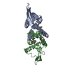
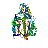
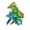
| Entry | Database: PDB / ID: 4hqe | ||||||
|---|---|---|---|---|---|---|---|
| Title | The crystal structure of QsrR-DNA complex | ||||||
 Components Components |
| ||||||
 Keywords Keywords | TRANSCRIPTION/DNA / Transcriptional regulator / DNA / TRANSCRIPTION-DNA complex | ||||||
| Function / homology |  Function and homology information Function and homology informationHelix-turn-helix, HxlR type / HxlR-like helix-turn-helix / HxlR-type HTH domain profile. / Winged helix-like DNA-binding domain superfamily/Winged helix DNA-binding domain / Arc Repressor Mutant, subunit A / Winged helix DNA-binding domain superfamily / Winged helix-like DNA-binding domain superfamily / Orthogonal Bundle / Mainly Alpha Similarity search - Domain/homology | ||||||
| Biological species |   Staphylococcus aureus (bacteria) Staphylococcus aureus (bacteria)synthetic construct (others) | ||||||
| Method |  X-RAY DIFFRACTION / SYNCHROTRON / MOLECULAR REPLACEMENT / Resolution: 2.299 Å X-RAY DIFFRACTION / SYNCHROTRON / MOLECULAR REPLACEMENT / Resolution: 2.299 Å | ||||||
 Authors Authors | He, C. / Ji, Q. / Zhang, L. | ||||||
 Citation Citation | Journal: Proc.Natl.Acad.Sci.USA / Year: 2013 Title: Molecular mechanism of quinone signaling mediated through S-quinonization of a YodB family repressor QsrR. Authors: Ji, Q. / Zhang, L. / Jones, M.B. / Sun, F. / Deng, X. / Liang, H. / Cho, H. / Brugarolas, P. / Gao, Y.N. / Peterson, S.N. / Lan, L. / Bae, T. / He, C. | ||||||
| History |
|
- Structure visualization
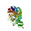
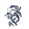
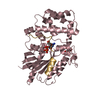
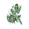
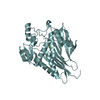
| Structure viewer | Molecule: MolmilJmol/JSmol |
|---|
- Downloads & links
-Download
| PDBx/mmCIF format | 4hqe.cif.gz | 76.9 KB | Display | PDBx/mmCIF format |
|---|---|---|---|---|
| PDB format | pdb4hqe.ent.gz | 54.8 KB | Display | PDB format |
| PDBx/mmJSON format | 4hqe.json.gz | Tree view | PDBx/mmJSON format | |
| Others |  Other downloads Other downloads |
-Validation report
| Summary document | 4hqe_validation.pdf.gz | 445.8 KB | Display | wwPDB validaton report |
|---|---|---|---|---|
| Full document | 4hqe_full_validation.pdf.gz | 449.7 KB | Display | |
| Data in XML | 4hqe_validation.xml.gz | 12 KB | Display | |
| Data in CIF | 4hqe_validation.cif.gz | 16.4 KB | Display | |
| Arichive directory | https://data.pdbj.org/pub/pdb/validation_reports/hq/4hqeftp://data.pdbj.org/pub/pdb/validation_reports/hq/4hqe | HTTPS FTP |
-Related structure data


-Links
 PDBj
PDBj


- Assembly
| Deposited unit | 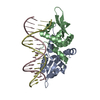
| ||||||||
|---|---|---|---|---|---|---|---|---|---|
| 1 |
| ||||||||
| Unit cell |
|
-Components
| #1: Protein | Mass: 13104.099 Da / Num. of mol.: 2 Source method: isolated from a genetically manipulated source Source: (gene. exp.) Staphylococcus aureus (bacteria) / Gene: SAV2123 / Production host: #2: DNA chain | | Mass: 5224.433 Da / Num. of mol.: 1 / Source method: obtained synthetically / Details: QsrR operator DNA1 / Source: (synth.) synthetic construct (others) #3: DNA chain | | Mass: 5184.409 Da / Num. of mol.: 1 / Source method: obtained synthetically / Details: QsrR operator DNA2 / Source: (synth.) synthetic construct (others) #4: Water | ChemComp-HOH / |  Mass: 18.015 Da / Num. of mol.: 97 / Source method: isolated from a natural source / Formula: H2O Mass: 18.015 Da / Num. of mol.: 97 / Source method: isolated from a natural source / Formula: H2O |
|---|
-Experimental details
-Experiment
| Experiment | Method: X-RAY DIFFRACTION / Number of used crystals: 1 |
|---|
- Sample preparation
| Crystal | Density Matthews: 2.49 Å3/Da / Density % sol: 50.52 % |
|---|---|
| Crystal grow | Temperature: 293 K / Method: vapor diffusion, sitting drop / pH: 8.5 Details: 0.2 M magnesium chloride, 0.1 M Tris.HCl, pH 8.5, 25% (w/v) polyethylene glycol 3350, VAPOR DIFFUSION, SITTING DROP, temperature 293K |
-Data collection
| Diffraction | Mean temperature: 100 K |
|---|---|
| Diffraction source | Source: SYNCHROTRON / Site: APS  / Beamline: 21-ID-D / Wavelength: 0.97872 Å / Beamline: 21-ID-D / Wavelength: 0.97872 Å |
| Detector | Type: MAR scanner 300 mm plate / Detector: IMAGE PLATE / Date: Mar 8, 2012 |
| Radiation | Protocol: SINGLE WAVELENGTH / Monochromatic (M) / Laue (L): M / Scattering type: x-ray |
| Radiation wavelength | Wavelength: 0.97872 Å / Relative weight: 1 |
| Reflection | Resolution: 2.299→25 Å / Num. obs: 15818 |
- Processing
| Software |
| |||||||||||||||||||||||||||||||||||||||||||||||||
|---|---|---|---|---|---|---|---|---|---|---|---|---|---|---|---|---|---|---|---|---|---|---|---|---|---|---|---|---|---|---|---|---|---|---|---|---|---|---|---|---|---|---|---|---|---|---|---|---|---|---|
| Refinement | Method to determine structure: MOLECULAR REPLACEMENT / Resolution: 2.299→23.815 Å / SU ML: 0.61 / σ(F): 1.36 / Phase error: 28.39 / Stereochemistry target values: ML
| |||||||||||||||||||||||||||||||||||||||||||||||||
| Solvent computation | Shrinkage radii: 0.86 Å / VDW probe radii: 1.1 Å / Solvent model: FLAT BULK SOLVENT MODEL / Bsol: 37.133 Å2 / ksol: 0.343 e/Å3 | |||||||||||||||||||||||||||||||||||||||||||||||||
| Displacement parameters |
| |||||||||||||||||||||||||||||||||||||||||||||||||
| Refinement step | Cycle: LAST / Resolution: 2.299→23.815 Å
| |||||||||||||||||||||||||||||||||||||||||||||||||
| Refine LS restraints |
| |||||||||||||||||||||||||||||||||||||||||||||||||
| LS refinement shell |
|
