 Movie
Movie Controller

[English] 日本語
 Yorodumi
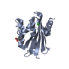
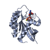
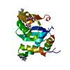
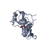
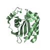
- PDB-4hfl: Crystal structure of the type VI effector Tae4 from Enterobacter ... -
+ Open data

- Basic information
| Entry | Database: PDB / ID: 4hfl | ||||||
|---|---|---|---|---|---|---|---|
| Title | Crystal structure of the type VI effector Tae4 from Enterobacter cloacae | ||||||
 Components Components | Putative cytoplasmic protein | ||||||
 Keywords Keywords | HYDROLASE / amidase | ||||||
| Function / homology | endopeptidase fold (from Nostoc punctiforme) - #70 / Type VI secretion system (T6SS), amidase effector protein 4 / Type VI secretion system (T6SS), amidase effector protein 4 / endopeptidase fold (from Nostoc punctiforme) / Alpha-Beta Complex / Alpha Beta / Putative cytoplasmic protein / Putative cytoplasmic protein Function and homology information Function and homology information | ||||||
| Biological species |  Enterobacter cloacae (bacteria) Enterobacter cloacae (bacteria) | ||||||
| Method |  X-RAY DIFFRACTION / SYNCHROTRON / MOLECULAR REPLACEMENT / Resolution: 2.002 Å X-RAY DIFFRACTION / SYNCHROTRON / MOLECULAR REPLACEMENT / Resolution: 2.002 Å | ||||||
 Authors Authors | Zhang, H. / Gao, Z.Q. / Dong, Y.H. | ||||||
 Citation Citation | Journal: J.Biol.Chem. / Year: 2013 Title: Structure of the type VI effector-immunity complex (Tae4-Tai4) provides novel insights into the inhibition mechanism of the effector by its immunity protein. Authors: Zhang, H. / Zhang, H. / Gao, Z.Q. / Wang, W.J. / Liu, G.F. / Xu, J.H. / Su, X.D. / Dong, Y.H. | ||||||
| History |
|
- Structure visualization
| Structure viewer | Molecule: MolmilJmol/JSmol |
|---|
- Downloads & links
-Download
| PDBx/mmCIF format | 4hfl.cif.gz | 79.1 KB | Display | PDBx/mmCIF format |
|---|---|---|---|---|
| PDB format | pdb4hfl.ent.gz | 59.2 KB | Display | PDB format |
| PDBx/mmJSON format | 4hfl.json.gz | Tree view | PDBx/mmJSON format | |
| Others |  Other downloads Other downloads |
-Validation report
| Arichive directory | https://data.pdbj.org/pub/pdb/validation_reports/hf/4hflftp://data.pdbj.org/pub/pdb/validation_reports/hf/4hfl | HTTPS FTP |
|---|
-Related structure data
| Related structure data |  4hffSC  4hfkC C: citing same article ( S: Starting model for refinement |
|---|---|
| Similar structure data |


-Links
 PDBj
PDBj- Assembly
| Deposited unit | 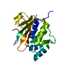
| ||||||||
|---|---|---|---|---|---|---|---|---|---|
| 1 |
| ||||||||
| 2 | 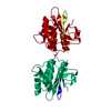
| ||||||||
| Unit cell |
|
-Components
| #1: Protein | Mass: 19165.059 Da / Num. of mol.: 1 Source method: isolated from a genetically manipulated source Source: (gene. exp.) Enterobacter cloacae (bacteria) / Strain: ATCC 13047 / DSM 30054 / NBRC 13535 / NCDC 279-56 / Gene: ECL_01542 / Production host: |
|---|---|
| #2: Water | ChemComp-HOH /  Mass: 18.015 Da / Num. of mol.: 98 / Source method: isolated from a natural source / Formula: H2O Mass: 18.015 Da / Num. of mol.: 98 / Source method: isolated from a natural source / Formula: H2O |
| Has protein modification | Y |
-Experimental details
-Experiment
| Experiment | Method: X-RAY DIFFRACTION / Number of used crystals: 1 |
|---|
- Sample preparation
| Crystal | Density Matthews: 2.32 Å3/Da / Density % sol: 47.09 % |
|---|---|
| Crystal grow | Temperature: 293 K / Method: vapor diffusion, sitting drop / pH: 5.6 Details: 0.5M Ammonium sulfate, 0.1M Sodium citrate tribasic dehydrate pH5.6, 1.0M Lithium sulfate monohydrate, VAPOR DIFFUSION, SITTING DROP, temperature 293K |
-Data collection
| Diffraction | Mean temperature: 100 K |
|---|---|
| Diffraction source | Source: SYNCHROTRON / Site: BSRF  / Beamline: 3W1A / Wavelength: 0.9793 Å / Beamline: 3W1A / Wavelength: 0.9793 Å |
| Detector | Type: MAR CCD 165 mm / Detector: CCD / Date: Jun 20, 2012 |
| Radiation | Monochromator: double crystal monochromator / Protocol: SINGLE WAVELENGTH / Monochromatic (M) / Laue (L): M / Scattering type: x-ray |
| Radiation wavelength | Wavelength: 0.9793 Å / Relative weight: 1 |
| Reflection | Resolution: 2→50 Å / Num. all: 12215 / Num. obs: 12215 / % possible obs: 100 % / Observed criterion σ(F): 0 / Observed criterion σ(I): 0 / Redundancy: 5.1 % / Rmerge(I) obs: 0.07 / Net I/σ(I): 44.3 |
| Reflection shell | Resolution: 2→2.03 Å / Redundancy: 4.8 % / Rmerge(I) obs: 0.142 / Mean I/σ(I) obs: 11.1 / Num. unique all: 593 / % possible all: 98.5 |
- Processing
| Software |
| ||||||||||||||||||||||||||||||||||||||||
|---|---|---|---|---|---|---|---|---|---|---|---|---|---|---|---|---|---|---|---|---|---|---|---|---|---|---|---|---|---|---|---|---|---|---|---|---|---|---|---|---|---|
| Refinement | Method to determine structure: MOLECULAR REPLACEMENT Starting model: PDB ENTRY 4HFF Resolution: 2.002→23.29 Å / SU ML: 0.26 / σ(F): 1.34 / Phase error: 24.74 / Stereochemistry target values: ML
| ||||||||||||||||||||||||||||||||||||||||
| Solvent computation | Shrinkage radii: 0.98 Å / VDW probe radii: 1.2 Å / Solvent model: FLAT BULK SOLVENT MODEL / Bsol: 48.544 Å2 / ksol: 0.366 e/Å3 | ||||||||||||||||||||||||||||||||||||||||
| Displacement parameters |
| ||||||||||||||||||||||||||||||||||||||||
| Refinement step | Cycle: LAST / Resolution: 2.002→23.29 Å
| ||||||||||||||||||||||||||||||||||||||||
| Refine LS restraints |
| ||||||||||||||||||||||||||||||||||||||||
| LS refinement shell | Refine-ID: X-RAY DIFFRACTION / Total num. of bins used: 4
| ||||||||||||||||||||||||||||||||||||||||
| Refinement TLS params. | Method: refined / Origin x: 4.2922 Å / Origin y: -33.864 Å / Origin z: 3.2202 Å
| ||||||||||||||||||||||||||||||||||||||||
| Refinement TLS group | Selection details: ALL |
