 Movie
Movie Controller


+ Open data

- Basic information
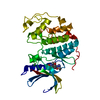
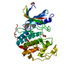
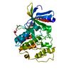
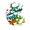
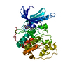
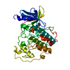
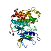
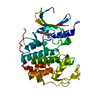
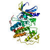
| Entry | Database: PDB / ID: 4ez3 | ||||||
|---|---|---|---|---|---|---|---|
| Title | CDK2 in complex with NSC 134199 | ||||||
 Components Components | Cyclin-dependent kinase 2 | ||||||
 Keywords Keywords | TRANSFERASE/TRANSFERASE INHIBITOR / protein kinase / TRANSFERASE-TRANSFERASE INHIBITOR complex | ||||||
| Function / homology |  Function and homology information Function and homology informationReplication initiator protein RctB, central region / RctB, helix turn helix domain / Vibrionales, replication initiator protein RctB, central region / RctB helix turn helix domain / Transferase(Phosphotransferase) domain 1 / Transferase(Phosphotransferase); domain 1 / Phosphorylase Kinase; domain 1 / Phosphorylase Kinase; domain 1 / Protein kinase domain / 2-Layer Sandwich ...Replication initiator protein RctB, central region / RctB, helix turn helix domain / Vibrionales, replication initiator protein RctB, central region / RctB helix turn helix domain / Transferase(Phosphotransferase) domain 1 / Transferase(Phosphotransferase); domain 1 / Phosphorylase Kinase; domain 1 / Phosphorylase Kinase; domain 1 / Protein kinase domain / 2-Layer Sandwich / Orthogonal Bundle / Mainly Alpha / Alpha Beta Similarity search - Domain/homology | ||||||
| Biological species |  Homo sapiens (human) Homo sapiens (human) | ||||||
| Method |  X-RAY DIFFRACTION / MOLECULAR REPLACEMENT / Resolution: 2 Å X-RAY DIFFRACTION / MOLECULAR REPLACEMENT / Resolution: 2 Å | ||||||
 Authors Authors | Alam, R. / Martin, M. / Zhu, J.-Y. / Schonbrunn, E. | ||||||
 Citation Citation | Journal: Chembiochem / Year: 2012 Title: A Novel Approach to the Discovery of Small-Molecule Ligands of CDK2. Authors: Martin, M.P. / Alam, R. / Betzi, S. / Ingles, D.J. / Zhu, J.Y. / Schonbrunn, E. | ||||||
| History |
|
- Structure visualization
| Structure viewer | Molecule: MolmilJmol/JSmol |
|---|
- Downloads & links
-Download
| PDBx/mmCIF format | 4ez3.cif.gz | 134 KB | Display | PDBx/mmCIF format |
|---|---|---|---|---|
| PDB format | pdb4ez3.ent.gz | 103.4 KB | Display | PDB format |
| PDBx/mmJSON format | 4ez3.json.gz | Tree view | PDBx/mmJSON format | |
| Others |  Other downloads Other downloads |
-Validation report
| Summary document | 4ez3_validation.pdf.gz | 692.4 KB | Display | wwPDB validaton report |
|---|---|---|---|---|
| Full document | 4ez3_full_validation.pdf.gz | 699.6 KB | Display | |
| Data in XML | 4ez3_validation.xml.gz | 14.6 KB | Display | |
| Data in CIF | 4ez3_validation.cif.gz | 19.8 KB | Display | |
| Arichive directory | https://data.pdbj.org/pub/pdb/validation_reports/ez/4ez3ftp://data.pdbj.org/pub/pdb/validation_reports/ez/4ez3 | HTTPS FTP |
-Related structure data
| Related structure data |  3ti1C 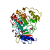 3tiyC 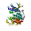 3tizC 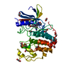 4erwC 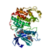 4ez7C  3pxzS C: citing same article ( S: Starting model for refinement |
|---|---|
| Similar structure data |


-Links
 PDBj
PDBj
- Assembly
| Deposited unit | 
| ||||||||
|---|---|---|---|---|---|---|---|---|---|
| 1 |
| ||||||||
| Unit cell |
|
-Components
| #1: Protein | Mass: 34761.348 Da / Num. of mol.: 1 Source method: isolated from a genetically manipulated source Source: (gene. exp.) Homo sapiens (human) / Gene: CDK2, CDKN2 / Plasmid: pGEX6P-1 / Production host:  |
|---|---|
| #2: Chemical | ChemComp-0S0 / 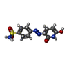  Mass: 294.287 Da / Num. of mol.: 1 / Source method: obtained synthetically / Formula: C11H10N4O4S Mass: 294.287 Da / Num. of mol.: 1 / Source method: obtained synthetically / Formula: C11H10N4O4S |
| #3: Chemical | ChemComp-EDO /   Mass: 62.068 Da / Num. of mol.: 1 / Source method: obtained synthetically / Formula: C2H6O2 Mass: 62.068 Da / Num. of mol.: 1 / Source method: obtained synthetically / Formula: C2H6O2 |
| #4: Water | ChemComp-HOH /  Mass: 18.015 Da / Num. of mol.: 97 / Source method: isolated from a natural source / Formula: H2O Mass: 18.015 Da / Num. of mol.: 97 / Source method: isolated from a natural source / Formula: H2O |
-Experimental details
-Experiment
| Experiment | Method: X-RAY DIFFRACTION / Number of used crystals: 1 |
|---|
- Sample preparation
| Crystal | Density Matthews: 1.93 Å3/Da / Density % sol: 36.35 % |
|---|---|
| Crystal grow | Temperature: 291 K / Method: vapor diffusion, hanging drop / pH: 7.5 Details: 5 mg/mL CDK2 protein, 5% v/v PEG3350, 50 mM HEPES/NaOH, pH 7.5, 50 mM sodium/potassium phosphate, pH 7.5, 10 mM NSC134199 soak, VAPOR DIFFUSION, HANGING DROP, temperature 291K |
-Data collection
| Diffraction | Mean temperature: 93 K |
|---|---|
| Diffraction source | Source: ROTATING ANODE / Type: RIGAKU MICROMAX-007 HF / Wavelength: 1.54178 |
| Detector | Type: RIGAKU SATURN 944+ / Detector: CCD / Date: Apr 29, 2012 |
| Radiation | Monochromator: MIRRORS / Protocol: SINGLE WAVELENGTH / Monochromatic (M) / Laue (L): M / Scattering type: x-ray |
| Radiation wavelength | Wavelength: 1.54178 Å / Relative weight: 1 |
| Reflection | Resolution: 2→20 Å / Num. obs: 18753 / % possible obs: 99.9 % / Observed criterion σ(I): -3 / Redundancy: 7 % / Rmerge(I) obs: 0.07 / Rsym value: 0.065 / Net I/σ(I): 21 |
| Reflection shell | Resolution: 2→2.1 Å / Redundancy: 7 % / Rmerge(I) obs: 0.359 / Mean I/σ(I) obs: 6.4 / Rsym value: 0.33 / % possible all: 99.9 |
- Processing
| Software |
| ||||||||||||||||||||||||||||||||||||||||||||||||||||||||
|---|---|---|---|---|---|---|---|---|---|---|---|---|---|---|---|---|---|---|---|---|---|---|---|---|---|---|---|---|---|---|---|---|---|---|---|---|---|---|---|---|---|---|---|---|---|---|---|---|---|---|---|---|---|---|---|---|---|
| Refinement | Method to determine structure: MOLECULAR REPLACEMENT Starting model: PDB ENTRY 3PXZ Resolution: 2→18.505 Å / SU ML: 0.2 / σ(F): 1.99 / Phase error: 21.78 / Stereochemistry target values: ML
| ||||||||||||||||||||||||||||||||||||||||||||||||||||||||
| Solvent computation | Shrinkage radii: 0.6 Å / VDW probe radii: 0.9 Å / Solvent model: FLAT BULK SOLVENT MODEL / Bsol: 60 Å2 / ksol: 0.4 e/Å3 | ||||||||||||||||||||||||||||||||||||||||||||||||||||||||
| Displacement parameters |
| ||||||||||||||||||||||||||||||||||||||||||||||||||||||||
| Refinement step | Cycle: LAST / Resolution: 2→18.505 Å
| ||||||||||||||||||||||||||||||||||||||||||||||||||||||||
| Refine LS restraints |
| ||||||||||||||||||||||||||||||||||||||||||||||||||||||||
| LS refinement shell |
|
