Protocol: SINGLE WAVELENGTH / Monochromatic (M) / Laue (L): M / Scattering type: x-ray
Radiation wavelength
Wavelength: 1.5418 Å / Relative weight: 1
Reflection
Resolution: 1.84→44.5 Å / Num. obs: 14005 / % possible obs: 97 % / Observed criterion σ(I): 0 / Redundancy: 3.1 % / Rmerge(I) obs: 0.06 / Net I/σ(I): 37
Reflection shell
Resolution: 1.84→1.9 Å / Redundancy: 2.3 % / Rmerge(I) obs: 0.1 / Mean I/σ(I) obs: 11 / % possible all: 85
-
Processing
Software
Name
Version
Classification
REFMAC
5.6.0117
refinement
HKL-2000
datareduction
SCALEPACK
datascaling
Refinement
Method to determine structure: OTHER Starting model: NONE Resolution: 1.84→44.55 Å / Cor.coef. Fo:Fc: 0.965 / Cor.coef. Fo:Fc free: 0.946 / SU B: 2.683 / SU ML: 0.083 / Cross valid method: THROUGHOUT / ESU R: 0.151 / ESU R Free: 0.135 / Stereochemistry target values: MAXIMUM LIKELIHOOD / Details: HYDROGENS HAVE BEEN USED IF PRESENT IN THE INPUT
Rfactor
Num. reflection
% reflection
Selection details
Rfree
0.1954
678
5 %
RANDOM
Rwork
0.14983
-
-
-
obs
0.15208
12950
97.38 %
-
Solvent computation
Ion probe radii: 0.8 Å / Shrinkage radii: 0.8 Å / VDW probe radii: 1.2 Å / Solvent model: MASK
 Movie
Movie Controller

 Yorodumi
 Open data
 Basic information
 Components
 Keywords
 Function and homology information
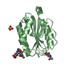
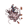
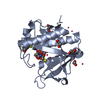
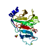
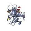
 STREPTOCOCCUS PNEUMONIAE (bacteria)
 X-RAY DIFFRACTION / OTHER / Resolution: 1.84 Å
  Authors
 Citation
 Structure visualization
 Downloads & links
 Other downloads
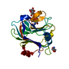


 PDBj
PDBj


 Assembly
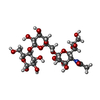


 Mass: 122.143 Da / Num. of mol.: 1 / Source method: obtained synthetically / Formula: C4H12NO3 / Comment: pH buffer*YM
 Mass: 18.015 Da / Num. of mol.: 148 / Source method: isolated from a natural source / Formula: H2O
 Sample preparation
 Processing