 Movie
Movie Controller


+ Open data

- Basic information
| Entry | Database: PDB / ID: 3x04 | ||||||
|---|---|---|---|---|---|---|---|
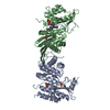
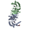
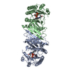
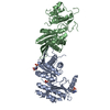
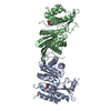
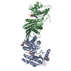
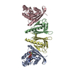
| Title | Crystal structure of PIP4KIIBETA complex with GMPPNP | ||||||
 Components Components | Phosphatidylinositol 5-phosphate 4-kinase type-2 beta | ||||||
 Keywords Keywords | TRANSFERASE / LIPID KINASE / PHOSPHOINOSITIDE SIGNALING | ||||||
| Function / homology |  Function and homology information Function and homology information1-phosphatidylinositol-5-phosphate 4-kinase / 1-phosphatidylinositol-5-phosphate 4-kinase activity / 1-phosphatidyl-1D-myo-inositol 4,5-bisphosphate biosynthetic process / Synthesis of PIPs in the nucleus / 1-phosphatidylinositol-4-phosphate 5-kinase activity / autophagosome-lysosome fusion / positive regulation of autophagosome assembly / phosphatidylinositol phosphate biosynthetic process / PI5P Regulates TP53 Acetylation / Synthesis of PIPs at the plasma membrane ...1-phosphatidylinositol-5-phosphate 4-kinase / 1-phosphatidylinositol-5-phosphate 4-kinase activity / 1-phosphatidyl-1D-myo-inositol 4,5-bisphosphate biosynthetic process / Synthesis of PIPs in the nucleus / 1-phosphatidylinositol-4-phosphate 5-kinase activity / autophagosome-lysosome fusion / positive regulation of autophagosome assembly / phosphatidylinositol phosphate biosynthetic process / PI5P Regulates TP53 Acetylation / Synthesis of PIPs at the plasma membrane / negative regulation of insulin receptor signaling pathway / autophagosome / regulation of autophagy / PI5P, PP2A and IER3 Regulate PI3K/AKT Signaling / cell surface receptor signaling pathway / endoplasmic reticulum membrane / GTP binding / protein homodimerization activity / nucleoplasm / ATP binding / nucleus / plasma membrane / cytosol Similarity search - Function | ||||||
| Biological species |  Homo sapiens (human) Homo sapiens (human) | ||||||
| Method |  X-RAY DIFFRACTION / SYNCHROTRON / MOLECULAR REPLACEMENT / Resolution: 2.6 Å X-RAY DIFFRACTION / SYNCHROTRON / MOLECULAR REPLACEMENT / Resolution: 2.6 Å | ||||||
 Authors Authors | Takeuchi, K. / Lo, Y.H. / Sumita, K. / Senda, M. / Terakawa, J. / Dimitoris, A. / Locasale, J.W. / Sasaki, M. / Yoshino, H. / Zhang, Y. ...Takeuchi, K. / Lo, Y.H. / Sumita, K. / Senda, M. / Terakawa, J. / Dimitoris, A. / Locasale, J.W. / Sasaki, M. / Yoshino, H. / Zhang, Y. / Kahoud, E.R. / Takano, T. / Yokota, T. / Emerling, B. / Asara, J.A. / Ishida, T. / Shimada, I. / Daikoku, T. / Cantley, L.C. / Senda, T. / Sasaki, A.T. | ||||||
 Citation Citation | Journal: Mol.Cell / Year: 2016 Title: The Lipid Kinase PI5P4K beta Is an Intracellular GTP Sensor for Metabolism and Tumorigenesis Authors: Sumita, K. / Lo, Y.H. / Takeuchi, K. / Senda, M. / Kofuji, S. / Ikeda, Y. / Terakawa, J. / Sasaki, M. / Yoshino, H. / Majd, N. / Zheng, Y. / Kahoud, E.R. / Yokota, T. / Emerling, B.M. / ...Authors: Sumita, K. / Lo, Y.H. / Takeuchi, K. / Senda, M. / Kofuji, S. / Ikeda, Y. / Terakawa, J. / Sasaki, M. / Yoshino, H. / Majd, N. / Zheng, Y. / Kahoud, E.R. / Yokota, T. / Emerling, B.M. / Asara, J.M. / Ishida, T. / Locasale, J.W. / Daikoku, T. / Anastasiou, D. / Senda, T. / Sasaki, A.T. | ||||||
| History |
|
- Structure visualization
| Structure viewer | Molecule: MolmilJmol/JSmol |
|---|
- Downloads & links
-Download
| PDBx/mmCIF format | 3x04.cif.gz | 275.1 KB | Display | PDBx/mmCIF format |
|---|---|---|---|---|
| PDB format | pdb3x04.ent.gz | 224.1 KB | Display | PDB format |
| PDBx/mmJSON format | 3x04.json.gz | Tree view | PDBx/mmJSON format | |
| Others |  Other downloads Other downloads |
-Validation report
| Arichive directory | https://data.pdbj.org/pub/pdb/validation_reports/x0/3x04ftp://data.pdbj.org/pub/pdb/validation_reports/x0/3x04 | HTTPS FTP |
|---|
-Related structure data
| Related structure data | 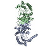 3wzzSC 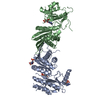 3x01C 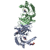 3x02C 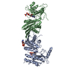 3x03C  3x05C  3x06C  3x07C 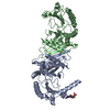 3x08C  3x09C 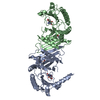 3x0aC 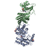 3x0bC 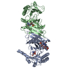 3x0cC S: Starting model for refinement C: citing same article ( |
|---|---|
| Similar structure data |


-Links
 PDBj
PDBj


- Assembly
| Deposited unit | 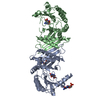
| ||||||||
|---|---|---|---|---|---|---|---|---|---|
| 1 |
| ||||||||
| Unit cell |
|
-Components
| #1: Protein | Mass: 44928.902 Da / Num. of mol.: 2 / Fragment: UNP RESIDUES 31-416 Source method: isolated from a genetically manipulated source Source: (gene. exp.) Homo sapiens (human) / Gene: PIP4K2B / Production host:  References: UniProt: P78356, 1-phosphatidylinositol-5-phosphate 4-kinase #2: Chemical | ChemComp-GNP /   Mass: 522.196 Da / Num. of mol.: 4 / Source method: obtained synthetically / Formula: C10H17N6O13P3 Mass: 522.196 Da / Num. of mol.: 4 / Source method: obtained synthetically / Formula: C10H17N6O13P3Comment: GppNHp, GMPPNP, energy-carrying molecule analogue*YM #3: Water | ChemComp-HOH / |  Mass: 18.015 Da / Num. of mol.: 17 / Source method: isolated from a natural source / Formula: H2O Mass: 18.015 Da / Num. of mol.: 17 / Source method: isolated from a natural source / Formula: H2O |
|---|
-Experimental details
-Experiment
| Experiment | Method: X-RAY DIFFRACTION / Number of used crystals: 1 |
|---|
- Sample preparation
| Crystal | Density Matthews: 3.01 Å3/Da / Density % sol: 59.16 % |
|---|---|
| Crystal grow | Temperature: 277 K / Method: vapor diffusion, sitting drop / pH: 5.5 Details: 100MM NA-CITRATE, 10MM MG-ACETATE, 100MM LI-ACETATE, 8-14%(V/V) PEG4000, pH 5.5, VAPOR DIFFUSION, SITTING DROP, temperature 277K |
-Data collection
| Diffraction | Mean temperature: 95 K |
|---|---|
| Diffraction source | Source: SYNCHROTRON / Site: Photon Factory  / Beamline: AR-NE3A / Wavelength: 1 / Wavelength: 1 Å / Beamline: AR-NE3A / Wavelength: 1 / Wavelength: 1 Å |
| Detector | Type: ADSC QUANTUM 270 / Detector: CCD / Date: Mar 11, 2012 |
| Radiation | Monochromator: SI111 / Protocol: SINGLE WAVELENGTH / Monochromatic (M) / Laue (L): M / Scattering type: x-ray |
| Radiation wavelength | Wavelength: 1 Å / Relative weight: 1 |
| Reflection | Resolution: 2.6→94.454 Å / Num. obs: 33027 / % possible obs: 97.9 % / Observed criterion σ(F): 0 / Observed criterion σ(I): 0 / Biso Wilson estimate: 70.82 Å2 |
| Reflection shell | Resolution: 2.6→2.74 Å / % possible all: 99.8 |
- Processing
| Software |
| |||||||||||||||||||||||||||||||||||||||||||||||||||||||||||||||||||||||||||||||||||||||||||||||||||||||||||||||||||||||||||||||||||||||||||||||||||||||||||||||||||||||||||||||||||||||||||||||||||||||||||||||||||||||||||||||||||||||||||||||||||||||||||||||||||||||||||||||||||
|---|---|---|---|---|---|---|---|---|---|---|---|---|---|---|---|---|---|---|---|---|---|---|---|---|---|---|---|---|---|---|---|---|---|---|---|---|---|---|---|---|---|---|---|---|---|---|---|---|---|---|---|---|---|---|---|---|---|---|---|---|---|---|---|---|---|---|---|---|---|---|---|---|---|---|---|---|---|---|---|---|---|---|---|---|---|---|---|---|---|---|---|---|---|---|---|---|---|---|---|---|---|---|---|---|---|---|---|---|---|---|---|---|---|---|---|---|---|---|---|---|---|---|---|---|---|---|---|---|---|---|---|---|---|---|---|---|---|---|---|---|---|---|---|---|---|---|---|---|---|---|---|---|---|---|---|---|---|---|---|---|---|---|---|---|---|---|---|---|---|---|---|---|---|---|---|---|---|---|---|---|---|---|---|---|---|---|---|---|---|---|---|---|---|---|---|---|---|---|---|---|---|---|---|---|---|---|---|---|---|---|---|---|---|---|---|---|---|---|---|---|---|---|---|---|---|---|---|---|---|---|---|---|---|---|---|---|---|---|---|---|---|---|---|---|---|---|---|---|---|---|---|---|---|---|---|---|---|---|---|---|---|---|---|---|---|---|---|---|---|---|---|---|---|---|---|---|
| Refinement | Method to determine structure: MOLECULAR REPLACEMENT Starting model: 3WZZ Resolution: 2.6→94.45 Å / SU ML: 0.56 / σ(F): 1.18 / Phase error: 26.98 / Stereochemistry target values: ML
| |||||||||||||||||||||||||||||||||||||||||||||||||||||||||||||||||||||||||||||||||||||||||||||||||||||||||||||||||||||||||||||||||||||||||||||||||||||||||||||||||||||||||||||||||||||||||||||||||||||||||||||||||||||||||||||||||||||||||||||||||||||||||||||||||||||||||||||||||||
| Solvent computation | Shrinkage radii: 0.98 Å / VDW probe radii: 1.2 Å / Solvent model: FLAT BULK SOLVENT MODEL / Bsol: 70.42 Å2 / ksol: 0.36 e/Å3 | |||||||||||||||||||||||||||||||||||||||||||||||||||||||||||||||||||||||||||||||||||||||||||||||||||||||||||||||||||||||||||||||||||||||||||||||||||||||||||||||||||||||||||||||||||||||||||||||||||||||||||||||||||||||||||||||||||||||||||||||||||||||||||||||||||||||||||||||||||
| Displacement parameters | Biso mean: 84.34 Å2
| |||||||||||||||||||||||||||||||||||||||||||||||||||||||||||||||||||||||||||||||||||||||||||||||||||||||||||||||||||||||||||||||||||||||||||||||||||||||||||||||||||||||||||||||||||||||||||||||||||||||||||||||||||||||||||||||||||||||||||||||||||||||||||||||||||||||||||||||||||
| Refinement step | Cycle: LAST / Resolution: 2.6→94.45 Å
| |||||||||||||||||||||||||||||||||||||||||||||||||||||||||||||||||||||||||||||||||||||||||||||||||||||||||||||||||||||||||||||||||||||||||||||||||||||||||||||||||||||||||||||||||||||||||||||||||||||||||||||||||||||||||||||||||||||||||||||||||||||||||||||||||||||||||||||||||||
| Refine LS restraints |
| |||||||||||||||||||||||||||||||||||||||||||||||||||||||||||||||||||||||||||||||||||||||||||||||||||||||||||||||||||||||||||||||||||||||||||||||||||||||||||||||||||||||||||||||||||||||||||||||||||||||||||||||||||||||||||||||||||||||||||||||||||||||||||||||||||||||||||||||||||
| LS refinement shell | Refine-ID: X-RAY DIFFRACTION / Total num. of bins used: 23
| |||||||||||||||||||||||||||||||||||||||||||||||||||||||||||||||||||||||||||||||||||||||||||||||||||||||||||||||||||||||||||||||||||||||||||||||||||||||||||||||||||||||||||||||||||||||||||||||||||||||||||||||||||||||||||||||||||||||||||||||||||||||||||||||||||||||||||||||||||
| Refinement TLS params. | Method: refined / Refine-ID: X-RAY DIFFRACTION
| |||||||||||||||||||||||||||||||||||||||||||||||||||||||||||||||||||||||||||||||||||||||||||||||||||||||||||||||||||||||||||||||||||||||||||||||||||||||||||||||||||||||||||||||||||||||||||||||||||||||||||||||||||||||||||||||||||||||||||||||||||||||||||||||||||||||||||||||||||
| Refinement TLS group |
|
