 Movie
Movie Controller


+ Open data

- Basic information
| Entry | Database: PDB / ID: 3w7z | ||||||
|---|---|---|---|---|---|---|---|
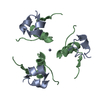
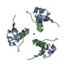
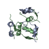
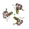
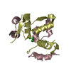
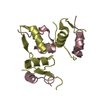
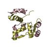
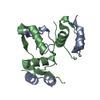
| Title | 1.15A structure of human 2Zn insulin at 293K | ||||||
 Components Components | (Insulin) x 2 | ||||||
 Keywords Keywords | HORMONE / Human insulin / glucose metabolism / secretion | ||||||
| Function / homology |  Function and homology information Function and homology informationnegative regulation of glycogen catabolic process / positive regulation of nitric oxide mediated signal transduction / negative regulation of fatty acid metabolic process / negative regulation of feeding behavior / Signaling by Insulin receptor / IRS activation / Insulin processing / regulation of protein secretion / positive regulation of peptide hormone secretion / positive regulation of respiratory burst ...negative regulation of glycogen catabolic process / positive regulation of nitric oxide mediated signal transduction / negative regulation of fatty acid metabolic process / negative regulation of feeding behavior / Signaling by Insulin receptor / IRS activation / Insulin processing / regulation of protein secretion / positive regulation of peptide hormone secretion / positive regulation of respiratory burst / Regulation of gene expression in beta cells / negative regulation of acute inflammatory response / alpha-beta T cell activation / Synthesis, secretion, and deacylation of Ghrelin / positive regulation of dendritic spine maintenance / negative regulation of protein secretion / negative regulation of gluconeogenesis / positive regulation of glycogen biosynthetic process / fatty acid homeostasis / Signal attenuation / positive regulation of insulin receptor signaling pathway / FOXO-mediated transcription of oxidative stress, metabolic and neuronal genes / negative regulation of respiratory burst involved in inflammatory response / negative regulation of lipid catabolic process / positive regulation of lipid biosynthetic process / negative regulation of oxidative stress-induced intrinsic apoptotic signaling pathway / regulation of protein localization to plasma membrane / transport vesicle / nitric oxide-cGMP-mediated signaling / positive regulation of nitric-oxide synthase activity / COPI-mediated anterograde transport / Insulin receptor recycling / negative regulation of reactive oxygen species biosynthetic process / positive regulation of brown fat cell differentiation / insulin-like growth factor receptor binding / NPAS4 regulates expression of target genes / neuron projection maintenance / endoplasmic reticulum-Golgi intermediate compartment membrane / positive regulation of mitotic nuclear division / Insulin receptor signalling cascade / positive regulation of glycolytic process / positive regulation of cytokine production / endosome lumen / acute-phase response / positive regulation of long-term synaptic potentiation / positive regulation of D-glucose import across plasma membrane / positive regulation of protein secretion / insulin receptor binding / positive regulation of cell differentiation / Regulation of insulin secretion / wound healing / positive regulation of neuron projection development / hormone activity / negative regulation of protein catabolic process / regulation of synaptic plasticity / positive regulation of protein localization to nucleus / Golgi lumen / vasodilation / cognition / glucose metabolic process / insulin receptor signaling pathway / cell-cell signaling / glucose homeostasis / regulation of protein localization / PI5P, PP2A and IER3 Regulate PI3K/AKT Signaling / positive regulation of cell growth / protease binding / secretory granule lumen / positive regulation of canonical NF-kappaB signal transduction / positive regulation of phosphatidylinositol 3-kinase/protein kinase B signal transduction / positive regulation of MAPK cascade / positive regulation of cell migration / G protein-coupled receptor signaling pathway / endoplasmic reticulum lumen / Amyloid fiber formation / receptor ligand activity / Golgi membrane / negative regulation of gene expression / positive regulation of cell population proliferation / positive regulation of gene expression / regulation of DNA-templated transcription / extracellular space / extracellular region / identical protein binding Similarity search - Function | ||||||
| Biological species |  Homo sapiens (human) Homo sapiens (human) | ||||||
| Method |  X-RAY DIFFRACTION / SYNCHROTRON / MOLECULAR REPLACEMENT / Resolution: 1.15 Å X-RAY DIFFRACTION / SYNCHROTRON / MOLECULAR REPLACEMENT / Resolution: 1.15 Å | ||||||
 Authors Authors | Hoshikawa, N. / Sasaki, K. / Sakabe, N. / Sakabe, K. | ||||||
 Citation Citation | Journal: To be Published Title: 1.15A structure of human 2Zn insulin at 293K Authors: Hoshikawa, N. / Sasaki, K. / Sakabe, N. / Sakabe, K. #1: Journal: NUCL INSTRUM METHODS PHYS RES A / Year: 2001Title: Automatic Weissenberg data collection system for time-resolved protein crystallography Authors: Sakabe, N. / Sakabe, K. / Higashi, T. / Igarashi, N. / Suzuki, M. / Watanabe, N. / Sasaki, K. #2: Journal: J.SYNCHROTRON RADIAT. / Year: 1999Title: Rotated-inclined focusing monochromator with simultaneous tuning of asymmetry factor and radius of curvature over a wide wavelength range Authors: Watanabe, N. / Suzuki, M. / Higashi, Y. / Sakabe, N. #3: Journal: J.SYNCHROTRON RADIAT. / Year: 1997Title: Large-Format Imaging Plate and Weissenberg Camera for Accurate Protein Crystallographic Data Collection Using Synchrotron Radiation Authors: Sakabe, K. / Sasaki, K. / Watanabe, N. / Suzuki, M. / Wang, Z.G. / Miyahara, J. / Sakabe, N. #4: Journal: PHILOS.TRANS.R.SOC.LOND.B BIOL / Year: 1988Title: The structure of 2Zn pig insulin crystals at 1.5A resolution Authors: Baker, E. / Blundell, T.L. / Cutfield, J.F. / Cutfield, S.M. / Dodson, E.J. / Dodson, G. / Hodgkin, D.M.C. / Hubbard, E. / Isaacs, N.W. / Reynolds, C.D. / Sakabe, K. / Sakabe, N. / Vijayan, N.M. #5: Journal: Acta Crystallogr.,Sect.A / Year: 1972Title: A statistical evaluation of absorption Authors: Katayama, C. / Sakabe, N. / Sakabe, K. #6: Journal: J.APPL.CRYSTALLOGR. / Year: 1989Title: The processing of diffraction data taken on a screenless Weissenberg camera for macromolecular crystallography Authors: Higashi, T. | ||||||
| History |
|
- Structure visualization
| Structure viewer | Molecule: MolmilJmol/JSmol |
|---|
- Downloads & links
-Download
| PDBx/mmCIF format | 3w7z.cif.gz | 65.5 KB | Display | PDBx/mmCIF format |
|---|---|---|---|---|
| PDB format | pdb3w7z.ent.gz | 49.1 KB | Display | PDB format |
| PDBx/mmJSON format | 3w7z.json.gz | Tree view | PDBx/mmJSON format | |
| Others |  Other downloads Other downloads |
-Validation report
| Arichive directory | https://data.pdbj.org/pub/pdb/validation_reports/w7/3w7zftp://data.pdbj.org/pub/pdb/validation_reports/w7/3w7z | HTTPS FTP |
|---|
-Related structure data
| Related structure data | 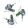 3w7yS S: Starting model for refinement |
|---|---|
| Similar structure data |


-Links
 PDBj
PDBj


- Assembly
| Deposited unit | 
| ||||||||||||
|---|---|---|---|---|---|---|---|---|---|---|---|---|---|
| 1 | 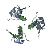
| ||||||||||||
| 2 | 
| ||||||||||||
| Unit cell |
| ||||||||||||
| Components on special symmetry positions |
|
-Components
| #1: Protein/peptide | Mass: 2383.698 Da / Num. of mol.: 2 / Fragment: UNP residues 90-110 / Source method: isolated from a natural source / Source: (natural) Homo sapiens (human) / References: UniProt: P01308#2: Protein/peptide | Mass: 3433.953 Da / Num. of mol.: 2 / Fragment: UNP residues 25-54 / Source method: isolated from a natural source / Source: (natural) Homo sapiens (human) / References: UniProt: P01308#3: Chemical |   Mass: 65.409 Da / Num. of mol.: 2 / Source method: obtained synthetically / Formula: Zn Mass: 65.409 Da / Num. of mol.: 2 / Source method: obtained synthetically / Formula: Zn#4: Water | ChemComp-HOH / |  Mass: 18.015 Da / Num. of mol.: 258 / Source method: isolated from a natural source / Formula: H2O Mass: 18.015 Da / Num. of mol.: 258 / Source method: isolated from a natural source / Formula: H2OHas protein modification | Y | |
|---|
-Experimental details
-Experiment
| Experiment | Method: X-RAY DIFFRACTION / Number of used crystals: 7 |
|---|
- Sample preparation
| Crystal | Density Matthews: 1.93 Å3/Da / Density % sol: 36.41 % |
|---|---|
| Crystal grow | Temperature: 293 K / Method: vapor diffusion, hanging drop / pH: 8.67 Details: 10mg/ml protein, 0.1M Sodium Citrate, 22%(v/v) DMF, 0.08%(w/v) Zinc chloride, pH 8.67, VAPOR DIFFUSION, HANGING DROP, temperature 293K |
-Data collection
| Diffraction | Mean temperature: 293 K |
|---|---|
| Diffraction source | Source: SYNCHROTRON / Site: Photon Factory  / Beamline: BL-6C / Wavelength: 1 Å / Beamline: BL-6C / Wavelength: 1 Å |
| Detector | Type: Fully automatic high speed Weissenberg data collection system called Galaxy Detector: IMAGE PLATE / Date: Apr 13, 2002 Details: The vertically focusing 1m long bent mirror of Pt-coated fused silica is 21m from the SR source point and 7m from the focus point |
| Radiation | Monochromator: Rotated-inclined focusing S(111) crystal / Protocol: SINGLE WAVELENGTH / Monochromatic (M) / Laue (L): M / Scattering type: x-ray |
| Radiation wavelength | Wavelength: 1 Å / Relative weight: 1 |
| Reflection | Resolution: 1→41.5 Å / Num. obs: 46687 / Redundancy: 8.4 % / Rmerge(I) obs: 0.066 |
- Processing
| Software |
| ||||||||||||||||||||||||||||||||||||||||||||||||||||||||||||
|---|---|---|---|---|---|---|---|---|---|---|---|---|---|---|---|---|---|---|---|---|---|---|---|---|---|---|---|---|---|---|---|---|---|---|---|---|---|---|---|---|---|---|---|---|---|---|---|---|---|---|---|---|---|---|---|---|---|---|---|---|---|
| Refinement | Method to determine structure: MOLECULAR REPLACEMENT Starting model: 3W7Y Resolution: 1.15→20 Å / Cor.coef. Fo:Fc: 0.976 / Cor.coef. Fo:Fc free: 0.957 / SU B: 1.286 / SU ML: 0.027 / Isotropic thermal model: anisotropic / Cross valid method: THROUGHOUT / ESU R: 0.04 / ESU R Free: 0.044 / Stereochemistry target values: MAXIMUM LIKELIHOOD
| ||||||||||||||||||||||||||||||||||||||||||||||||||||||||||||
| Solvent computation | Ion probe radii: 0.8 Å / Shrinkage radii: 0.8 Å / VDW probe radii: 1.2 Å / Solvent model: BABINET MODEL WITH MASK | ||||||||||||||||||||||||||||||||||||||||||||||||||||||||||||
| Displacement parameters | Biso mean: 27.135 Å2
| ||||||||||||||||||||||||||||||||||||||||||||||||||||||||||||
| Refinement step | Cycle: LAST / Resolution: 1.15→20 Å
| ||||||||||||||||||||||||||||||||||||||||||||||||||||||||||||
| Refine LS restraints |
| ||||||||||||||||||||||||||||||||||||||||||||||||||||||||||||
| LS refinement shell | Resolution: 1.15→1.18 Å / Total num. of bins used: 20
|
