 Movie
Movie Controller

[English] 日本語
 Yorodumi
- PDB-3upp: Structure of penicillin-binding protein A from M. tuberculosis: c... -
+ Open data

- Basic information
| Entry | Database: PDB / ID: 3upp | |||||||||
|---|---|---|---|---|---|---|---|---|---|---|
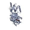
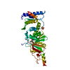
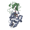
| Title | Structure of penicillin-binding protein A from M. tuberculosis: ceftrixaone acyl-enzyme complex | |||||||||
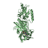
 Components Components | Penicillin-binding protein A | |||||||||
 Keywords Keywords | Penicillin-binding protein/Antibiotic / TRANSPEPTIDASE / peptidoglycan / beta-lactam / Penicillin-binding protein-Antibiotic complex | |||||||||
| Function / homology |  Function and homology information Function and homology informationpeptidoglycan L,D-transpeptidase activity / serine-type D-Ala-D-Ala carboxypeptidase / serine-type D-Ala-D-Ala carboxypeptidase activity / penicillin binding / membrane => GO:0016020 / peptidoglycan biosynthetic process / cell wall organization / regulation of cell shape / plasma membrane / cytosol Similarity search - Function | |||||||||
| Biological species |   Mycobacterium tuberculosis (bacteria) Mycobacterium tuberculosis (bacteria) | |||||||||
| Method |  X-RAY DIFFRACTION / SYNCHROTRON / FOURIER SYNTHESIS / Resolution: 2.4 Å X-RAY DIFFRACTION / SYNCHROTRON / FOURIER SYNTHESIS / Resolution: 2.4 Å | |||||||||
 Authors Authors | Davies, C. / Fedorovich, A. | |||||||||
 Citation Citation | Journal: J.Mol.Biol. / Year: 2012 Title: The role of the beta5-alpha11 loop in the active-site dynamics of acylated penicillin-binding protein A from Mycobacterium tuberculosis Authors: Fedarovich, A. / Nicholas, R.A. / Davies, C. | |||||||||
| History |
|
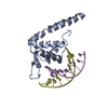
- Structure visualization
| Structure viewer | Molecule: MolmilJmol/JSmol |
|---|
- Downloads & links
-Download
| PDBx/mmCIF format | 3upp.cif.gz | 329 KB | Display | PDBx/mmCIF format |
|---|---|---|---|---|
| PDB format | pdb3upp.ent.gz | 270.1 KB | Display | PDB format |
| PDBx/mmJSON format | 3upp.json.gz | Tree view | PDBx/mmJSON format | |
| Others |  Other downloads Other downloads |
-Validation report
| Arichive directory | https://data.pdbj.org/pub/pdb/validation_reports/up/3uppftp://data.pdbj.org/pub/pdb/validation_reports/up/3upp | HTTPS FTP |
|---|
-Related structure data
| Related structure data | 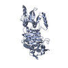 3un7C 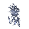 3upnC  3upoC 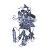 3lo7S S: Starting model for refinement C: citing same article ( |
|---|---|
| Similar structure data |


-Links
 PDBj
PDBj
- Assembly
| Deposited unit | 
| ||||||||
|---|---|---|---|---|---|---|---|---|---|
| 1 | 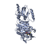
| ||||||||
| 2 | 
| ||||||||
| Unit cell |
|
-Components
| #1: Protein | Mass: 48464.492 Da / Num. of mol.: 2 / Fragment: UNP residues 35-491 Source method: isolated from a genetically manipulated source Source: (gene. exp.) Mycobacterium tuberculosis (bacteria) / Strain: H37Rv / Gene: MT0019, MTCY10H4.16c, pbpA, Rv0016c / Plasmid: pT7HTb / Production host: References: UniProt: P71586, UniProt: P9WKD1*PLUS, serine-type D-Ala-D-Ala carboxypeptidase #2: Chemical | 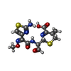  Mass: 397.429 Da / Num. of mol.: 2 / Source method: obtained synthetically / Formula: C14H15N5O5S2 / Comment: antibiotic*YM Mass: 397.429 Da / Num. of mol.: 2 / Source method: obtained synthetically / Formula: C14H15N5O5S2 / Comment: antibiotic*YM#3: Water | ChemComp-HOH / |  Mass: 18.015 Da / Num. of mol.: 21 / Source method: isolated from a natural source / Formula: H2O Mass: 18.015 Da / Num. of mol.: 21 / Source method: isolated from a natural source / Formula: H2OHas protein modification | Y | Nonpolymer details | THE STARTING ANTIBIOTIC | Sequence details | THE MUTATION G384R OCCURED DURING PCR | |
|---|
-Experimental details
-Experiment
| Experiment | Method: X-RAY DIFFRACTION / Number of used crystals: 1 |
|---|
- Sample preparation
| Crystal | Density Matthews: 2.29 Å3/Da / Density % sol: 46.19 % |
|---|---|
| Crystal grow | Temperature: 292 K / Method: vapor diffusion, hanging drop / pH: 5.5 Details: 25% PEG 3350, 0.2 M NaCl, 0.1 M Bis-Tris pH 5.5, VAPOR DIFFUSION, HANGING DROP, temperature 292K |
-Data collection
| Diffraction | Mean temperature: 100 K |
|---|---|
| Diffraction source | Source: SYNCHROTRON / Site: APS  / Beamline: 22-ID / Wavelength: 1 Å / Beamline: 22-ID / Wavelength: 1 Å |
| Detector | Type: MARMOSAIC 300 mm CCD / Detector: CCD / Date: Jun 12, 2011 |
| Radiation | Monochromator: SI(220) / Protocol: SINGLE WAVELENGTH / Monochromatic (M) / Laue (L): M / Scattering type: x-ray |
| Radiation wavelength | Wavelength: 1 Å / Relative weight: 1 |
| Reflection | Resolution: 2.4→45.7 Å / Num. all: 33749 / Num. obs: 33749 / % possible obs: 98.7 % / Observed criterion σ(F): 0 / Observed criterion σ(I): 0 / Redundancy: 10.2 % / Rmerge(I) obs: 0.08 / Net I/σ(I): 34.5 |
| Reflection shell | Resolution: 2.4→2.49 Å / Redundancy: 7.3 % / Rmerge(I) obs: 0.623 / Mean I/σ(I) obs: 2.2 / % possible all: 89.6 |
- Processing
| Software |
| ||||||||||||||||||||||||||||||||||||||||||||||||||||||||||||||||||||||||||||||||||||||||||||||||||||||||||||||||||||||||||||||||||||||||||||||||||||||||||||||||||||||||||
|---|---|---|---|---|---|---|---|---|---|---|---|---|---|---|---|---|---|---|---|---|---|---|---|---|---|---|---|---|---|---|---|---|---|---|---|---|---|---|---|---|---|---|---|---|---|---|---|---|---|---|---|---|---|---|---|---|---|---|---|---|---|---|---|---|---|---|---|---|---|---|---|---|---|---|---|---|---|---|---|---|---|---|---|---|---|---|---|---|---|---|---|---|---|---|---|---|---|---|---|---|---|---|---|---|---|---|---|---|---|---|---|---|---|---|---|---|---|---|---|---|---|---|---|---|---|---|---|---|---|---|---|---|---|---|---|---|---|---|---|---|---|---|---|---|---|---|---|---|---|---|---|---|---|---|---|---|---|---|---|---|---|---|---|---|---|---|---|---|---|---|---|
| Refinement | Method to determine structure: FOURIER SYNTHESIS Starting model: PDB entry 3LO7 Resolution: 2.4→40.29 Å / Cor.coef. Fo:Fc: 0.953 / Cor.coef. Fo:Fc free: 0.935 / SU B: 20.76 / SU ML: 0.223 / Isotropic thermal model: isotropic / Cross valid method: THROUGHOUT / ESU R: 0.531 / ESU R Free: 0.275 / Stereochemistry target values: MAXIMUM LIKELIHOOD / Details: HYDROGENS HAVE BEEN ADDED IN THE RIDING POSITIONS
| ||||||||||||||||||||||||||||||||||||||||||||||||||||||||||||||||||||||||||||||||||||||||||||||||||||||||||||||||||||||||||||||||||||||||||||||||||||||||||||||||||||||||||
| Solvent computation | Ion probe radii: 0.8 Å / Shrinkage radii: 0.8 Å / VDW probe radii: 1.2 Å / Solvent model: MASK | ||||||||||||||||||||||||||||||||||||||||||||||||||||||||||||||||||||||||||||||||||||||||||||||||||||||||||||||||||||||||||||||||||||||||||||||||||||||||||||||||||||||||||
| Displacement parameters | Biso mean: 52.992 Å2
| ||||||||||||||||||||||||||||||||||||||||||||||||||||||||||||||||||||||||||||||||||||||||||||||||||||||||||||||||||||||||||||||||||||||||||||||||||||||||||||||||||||||||||
| Refinement step | Cycle: LAST / Resolution: 2.4→40.29 Å
| ||||||||||||||||||||||||||||||||||||||||||||||||||||||||||||||||||||||||||||||||||||||||||||||||||||||||||||||||||||||||||||||||||||||||||||||||||||||||||||||||||||||||||
| Refine LS restraints |
| ||||||||||||||||||||||||||||||||||||||||||||||||||||||||||||||||||||||||||||||||||||||||||||||||||||||||||||||||||||||||||||||||||||||||||||||||||||||||||||||||||||||||||
| LS refinement shell | Resolution: 2.4→2.462 Å / Total num. of bins used: 20
|
