 Movie
Movie Controller

[English] 日本語
 Yorodumi
- PDB-3tgl: STRUCTURE AND MOLECULAR MODEL REFINEMENT OF RHIZOMUCOR MIEHEI TRI... -
+ Open data

- Basic information
| Entry | Database: PDB / ID: 3tgl | ||||||
|---|---|---|---|---|---|---|---|
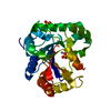
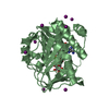
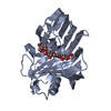
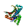
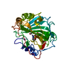
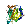
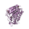
| Title | STRUCTURE AND MOLECULAR MODEL REFINEMENT OF RHIZOMUCOR MIEHEI TRIACYLGLYCERIDE LIPASE: A CASE STUDY OF THE USE OF SIMULATED ANNEALING IN PARTIAL MODEL REFINEMENT | ||||||
 Components Components | TRIACYL-GLYCEROL ACYLHYDROLASE | ||||||
 Keywords Keywords | HYDROLASE(CARBOXYLIC ESTERASE) | ||||||
| Function / homology |  Function and homology information Function and homology informationtriacylglycerol lipase / triacylglycerol lipase activity / lipid catabolic process / metal ion binding Similarity search - Function | ||||||
| Biological species |  Rhizomucor miehei (fungus) Rhizomucor miehei (fungus) | ||||||
| Method |  X-RAY DIFFRACTION / Resolution: 1.9 Å X-RAY DIFFRACTION / Resolution: 1.9 Å | ||||||
 Authors Authors | Brady, L. / Brzozowski, A.M. / Derewenda, Z.S. / Dodson, E.J. / Dodson, G.G. / Tolley, S.P. / Turkenburg, J.P. / Christiansen, L. / Huge-Jensen, B. / Norskov, L. / Thim, L. | ||||||
 Citation Citation | Journal: Acta Crystallogr.,Sect.B / Year: 1992 Title: STRUCTURE AND MOLECULAR-MODEL REFINEMENT OF RHIZOMUCOR-MIEHEI TRIACYLGLYCERIDE LIPASE - A CASE-STUDY OF THE USE OF SIMULATED ANNEALING IN PARTIAL MODEL REFINEMENT. Authors: Brzozowski, A.M. / Derewenda, Z.S. / Dodson, E.J. / Dodson, G.G. / Turkenburg, J.P. #1: Journal: J.Mol.Biol. / Year: 1992Title: The Crystal and Molecular Structure of the Rhizomucor Miehei Triacylglyceride Lipase at 1.9 Angstroms Resolution Authors: Derewenda, Z.S. / Derewenda, U. / Dodson, G.G. #2: Journal: Nature / Year: 1991Title: A Model for Interfacial Activation in Lipases from the Structure of a Fungal Lipase-Inhibitor Complex Authors: Brzozowski, A.M. / Derewenda, U. / Derewenda, Z.S. / Dodson, G.G. / Lawson, D.M. / Turkenburg, J.P. / Bjorkling, F. / Huge-Jensen, B. / Patka, S.A. / Thim, L. #3: Journal: Nature / Year: 1990Title: A Serine Protease Triad Forms the Catalytic Centre of a Triacylglycerol Lipase Authors: Brady, L. / Brzozowski, A.M. / Derewenda, Z.S. / Dodson, E. / Dodson, G. / Tolley, S. / Turkenburg, J.P. / Christiansen, L. / Huge-Jensen, B. / Norskov, L. / Thim, L. / Menge, U. | ||||||
| History |
|
- Structure visualization
| Structure viewer | Molecule: MolmilJmol/JSmol |
|---|
- Downloads & links
-Download
| PDBx/mmCIF format | 3tgl.cif.gz | 71.1 KB | Display | PDBx/mmCIF format |
|---|---|---|---|---|
| PDB format | pdb3tgl.ent.gz | 49.6 KB | Display | PDB format |
| PDBx/mmJSON format | 3tgl.json.gz | Tree view | PDBx/mmJSON format | |
| Others |  Other downloads Other downloads |
-Validation report
| Arichive directory | https://data.pdbj.org/pub/pdb/validation_reports/tg/3tglftp://data.pdbj.org/pub/pdb/validation_reports/tg/3tgl | HTTPS FTP |
|---|
-Related structure data
| Similar structure data |
|---|


-Links
 PDBj
PDBj- Assembly
| Deposited unit | 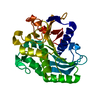
| ||||||||
|---|---|---|---|---|---|---|---|---|---|
| 1 |
| ||||||||
| Unit cell |
| ||||||||
| Atom site foot note | 1: RESIDUES PRO 34, PRO 209, PRO 229, AND PRO 250 ARE CIS PROLINES. |
-Components
| #1: Protein | Mass: 29594.043 Da / Num. of mol.: 1 Source method: isolated from a genetically manipulated source Source: (gene. exp.) Rhizomucor miehei (fungus) / References: UniProt: P19515, triacylglycerol lipase |
|---|---|
| #2: Water | ChemComp-HOH /  Mass: 18.015 Da / Num. of mol.: 230 / Source method: isolated from a natural source / Formula: H2O Mass: 18.015 Da / Num. of mol.: 230 / Source method: isolated from a natural source / Formula: H2O |
| Has protein modification | Y |
| Sequence details | RESIDUE 156 IN THIS ENTRY IS ASP AS IDENTIFIED BY ELECTRON DENSITY. IN THE CDNA SEQUENCE IT WAS ...RESIDUE 156 IN THIS ENTRY IS ASP AS IDENTIFIED |
-Experimental details
-Experiment
| Experiment | Method: X-RAY DIFFRACTION |
|---|
- Sample preparation
| Crystal | Density Matthews: 2.49 Å3/Da / Density % sol: 50.68 % | ||||||||||||||||||||
|---|---|---|---|---|---|---|---|---|---|---|---|---|---|---|---|---|---|---|---|---|---|
| Crystal grow | *PLUS pH: 8.05 / Method: vapor diffusion, hanging drop | ||||||||||||||||||||
| Components of the solutions | *PLUS
|
-Data collection
| Reflection | *PLUS Highest resolution: 1.9 Å / Lowest resolution: 3.45 Å / Num. all: 24101 / % possible obs: 82.8 % / Observed criterion σ(I): 2 / Redundancy: 5.5 % / Rmerge F obs: 0.0807 |
|---|
- Processing
| Software | Name: PROLSQ / Classification: refinement | ||||||||||||||||||||||||||||||||||||||||||||||||||||||||||||||||||||||||||||||||||||
|---|---|---|---|---|---|---|---|---|---|---|---|---|---|---|---|---|---|---|---|---|---|---|---|---|---|---|---|---|---|---|---|---|---|---|---|---|---|---|---|---|---|---|---|---|---|---|---|---|---|---|---|---|---|---|---|---|---|---|---|---|---|---|---|---|---|---|---|---|---|---|---|---|---|---|---|---|---|---|---|---|---|---|---|---|---|
| Refinement | Resolution: 1.9→7.5 Å / Rfactor obs: 0.129 Details: THERE ARE SOME ERRORS IN THE SEQUENCE DUE TO UNCERTAINTY IN THE ORIENTATION OF SIDE CHAINS. THESE DIFFERENCES ARE MINOR AND IN NO WAY COMPROMISE THE OVERALL QUALITY OF THE STRUCTURE OR THE ...Details: THERE ARE SOME ERRORS IN THE SEQUENCE DUE TO UNCERTAINTY IN THE ORIENTATION OF SIDE CHAINS. THESE DIFFERENCES ARE MINOR AND IN NO WAY COMPROMISE THE OVERALL QUALITY OF THE STRUCTURE OR THE SUITABILITY FOR MODELING OR MOLECULAR REPLACEMENT. SEQUENCE ADVISORY NOTICE: DIFFERENCE BETWEEN PIR AND PDB SEQUENCE. PIR ENTRY NAME: A34959 PIR RESIDUE PDB SEQRES NAME NUMBER NAME CHAIN SEQ/INSERT CODE ALA 150 VAL 150 GLY 156 ASP 156 | ||||||||||||||||||||||||||||||||||||||||||||||||||||||||||||||||||||||||||||||||||||
| Refinement step | Cycle: LAST / Resolution: 1.9→7.5 Å
| ||||||||||||||||||||||||||||||||||||||||||||||||||||||||||||||||||||||||||||||||||||
| Refine LS restraints |
| ||||||||||||||||||||||||||||||||||||||||||||||||||||||||||||||||||||||||||||||||||||
| Refinement | *PLUS Highest resolution: 1.9 Å / Lowest resolution: 7.5 Å / Num. reflection obs: 18960 / σ(F): 2 / Rfactor obs: 0.129 | ||||||||||||||||||||||||||||||||||||||||||||||||||||||||||||||||||||||||||||||||||||
| Solvent computation | *PLUS | ||||||||||||||||||||||||||||||||||||||||||||||||||||||||||||||||||||||||||||||||||||
| Displacement parameters | *PLUS |
