 Movie
Movie Controller

[English] 日本語
 Yorodumi
- PDB-3t2b: Fructose-1,6-bisphosphate aldolase/phosphatase from Thermoproteus... -
+ Open data

- Basic information
| Entry | Database: PDB / ID: 3t2b | ||||||
|---|---|---|---|---|---|---|---|
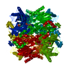
| Title | Fructose-1,6-bisphosphate aldolase/phosphatase from Thermoproteus neutrophilus, ligand free | ||||||
 Components Components | Fructose-1,6-bisphosphate aldolase/phosphatase | ||||||
 Keywords Keywords | LYASE / HYDROLASE / (beta/alpha)8 TIM barrel / FBP / F6P / DHAP / GAP / phosphorylation | ||||||
| Function / homology |  Function and homology information Function and homology informationfructose-bisphosphate aldolase / fructose-bisphosphate aldolase activity / fructose-bisphosphatase / fructose 1,6-bisphosphate 1-phosphatase activity / gluconeogenesis / magnesium ion binding Similarity search - Function | ||||||
| Biological species |   Thermoproteus neutrophilus (archaea) Thermoproteus neutrophilus (archaea) | ||||||
| Method |  X-RAY DIFFRACTION / SYNCHROTRON / MOLECULAR REPLACEMENT / Resolution: 1.52 Å X-RAY DIFFRACTION / SYNCHROTRON / MOLECULAR REPLACEMENT / Resolution: 1.52 Å | ||||||
 Authors Authors | Du, J. / Say, R. / Lue, W. / Fuchs, G. / Einsle, O. | ||||||
 Citation Citation | Journal: Nature / Year: 2011 Title: Active-site remodelling in the bifunctional fructose-1,6-bisphosphate aldolase/phosphatase. Authors: Du, J. / Say, R.F. / Lu, W. / Fuchs, G. / Einsle, O. | ||||||
| History |
|
- Structure visualization
| Structure viewer | Molecule: MolmilJmol/JSmol |
|---|
- Downloads & links
-Download
| PDBx/mmCIF format | 3t2b.cif.gz | 97.2 KB | Display | PDBx/mmCIF format |
|---|---|---|---|---|
| PDB format | pdb3t2b.ent.gz | 73.4 KB | Display | PDB format |
| PDBx/mmJSON format | 3t2b.json.gz | Tree view | PDBx/mmJSON format | |
| Others |  Other downloads Other downloads |
-Validation report
| Summary document | 3t2b_validation.pdf.gz | 431.9 KB | Display | wwPDB validaton report |
|---|---|---|---|---|
| Full document | 3t2b_full_validation.pdf.gz | 441.1 KB | Display | |
| Data in XML | 3t2b_validation.xml.gz | 19.9 KB | Display | |
| Data in CIF | 3t2b_validation.cif.gz | 29.8 KB | Display | |
| Arichive directory | https://data.pdbj.org/pub/pdb/validation_reports/t2/3t2bftp://data.pdbj.org/pub/pdb/validation_reports/t2/3t2b | HTTPS FTP |
-Related structure data
| Related structure data |  3t2cC 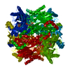 3t2dC  3t2eC 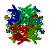 3t2fC 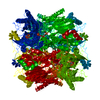 3t2gC  1umgS C: citing same article ( S: Starting model for refinement |
|---|---|
| Similar structure data |


-Links
 PDBj
PDBj
- Assembly
| Deposited unit | 
| ||||||||||||
|---|---|---|---|---|---|---|---|---|---|---|---|---|---|
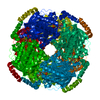
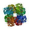
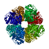
| 1 | x 8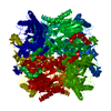
| ||||||||||||
| Unit cell |
| ||||||||||||
| Components on special symmetry positions |
|
-Components
| #1: Protein | Mass: 45461.031 Da / Num. of mol.: 1 Source method: isolated from a genetically manipulated source Source: (gene. exp.) Thermoproteus neutrophilus (archaea) / Strain: DSM 2338 / JCM 9278 / V24Sta / Gene: Tneu_0133 / Plasmid: pET23b / Production host:  References: UniProt: B1YAL1, fructose-bisphosphate aldolase, fructose-bisphosphatase | ||
|---|---|---|---|
| #2: Chemical |   Mass: 24.305 Da / Num. of mol.: 2 / Source method: obtained synthetically / Formula: Mg Mass: 24.305 Da / Num. of mol.: 2 / Source method: obtained synthetically / Formula: Mg#3: Water | ChemComp-HOH / |  Mass: 18.015 Da / Num. of mol.: 320 / Source method: isolated from a natural source / Formula: H2O Mass: 18.015 Da / Num. of mol.: 320 / Source method: isolated from a natural source / Formula: H2O |
-Experimental details
-Experiment
| Experiment | Method: X-RAY DIFFRACTION / Number of used crystals: 1 |
|---|
- Sample preparation
| Crystal | Density Matthews: 2.63 Å3/Da / Density % sol: 53.25 % |
|---|---|
| Crystal grow | Temperature: 294 K / Method: vapor diffusion, hanging drop / pH: 7.5 Details: 8% PEG3350, 0.1 M HEPES/NaOH, pH 7.5, VAPOR DIFFUSION, HANGING DROP, temperature 294K |
-Data collection
| Diffraction | Mean temperature: 90 K | |||||||||
|---|---|---|---|---|---|---|---|---|---|---|
| Diffraction source | Source: SYNCHROTRON / Site: SLS  / Beamline: X06SA / Wavelength: 1 / Wavelength: 1 Å / Beamline: X06SA / Wavelength: 1 / Wavelength: 1 Å | |||||||||
| Detector | Type: PSI PILATUS 6M / Detector: PIXEL / Date: Aug 1, 2010 | |||||||||
| Radiation | Monochromator: Fixed-exit LN2 cooled Double Crystal / Protocol: SINGLE WAVELENGTH / Monochromatic (M) / Laue (L): M / Scattering type: x-ray | |||||||||
| Radiation wavelength |
| |||||||||
| Reflection | Resolution: 1.52→54.794 Å / Num. all: 74360 / Num. obs: 74283 / % possible obs: 99.9 % / Observed criterion σ(F): 2 / Observed criterion σ(I): 2 / Redundancy: 10 % / Rmerge(I) obs: 0.064 / Rsym value: 0.064 / Net I/σ(I): 22.4 | |||||||||
| Reflection shell | Resolution: 1.52→1.62 Å / Redundancy: 10.3 % / Rmerge(I) obs: 0.363 / Mean I/σ(I) obs: 6 / Rsym value: 0.363 / % possible all: 99.9 |
- Processing
| Software |
| |||||||||||||||||||||||||||||||||||||||||||||||||||||||||||||||||
|---|---|---|---|---|---|---|---|---|---|---|---|---|---|---|---|---|---|---|---|---|---|---|---|---|---|---|---|---|---|---|---|---|---|---|---|---|---|---|---|---|---|---|---|---|---|---|---|---|---|---|---|---|---|---|---|---|---|---|---|---|---|---|---|---|---|---|
| Refinement | Method to determine structure: MOLECULAR REPLACEMENT Starting model: PDB ENTRY 1UMG Resolution: 1.52→54.79 Å / Cor.coef. Fo:Fc: 0.967 / Cor.coef. Fo:Fc free: 0.958 / SU B: 1.085 / SU ML: 0.04 / Cross valid method: THROUGHOUT / σ(F): 2 / ESU R: 0.062 / ESU R Free: 0.064 / Stereochemistry target values: MAXIMUM LIKELIHOOD Details: HYDROGENS HAVE BEEN ADDED IN THE RIDING POSITIONS U VALUES : REFINED INDIVIDUALLY
| |||||||||||||||||||||||||||||||||||||||||||||||||||||||||||||||||
| Solvent computation | Ion probe radii: 0.8 Å / Shrinkage radii: 0.8 Å / VDW probe radii: 1.4 Å / Solvent model: MASK | |||||||||||||||||||||||||||||||||||||||||||||||||||||||||||||||||
| Displacement parameters | Biso mean: 19.389 Å2
| |||||||||||||||||||||||||||||||||||||||||||||||||||||||||||||||||
| Refinement step | Cycle: LAST / Resolution: 1.52→54.79 Å
| |||||||||||||||||||||||||||||||||||||||||||||||||||||||||||||||||
| Refine LS restraints |
| |||||||||||||||||||||||||||||||||||||||||||||||||||||||||||||||||
| LS refinement shell | Resolution: 1.52→1.559 Å / Total num. of bins used: 20
|
