 Movie
Movie Controller


+ Open data

- Basic information
| Entry | Database: PDB / ID: 3ogb | ||||||
|---|---|---|---|---|---|---|---|
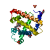
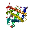
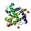
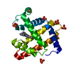
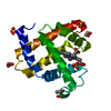
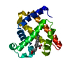
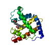
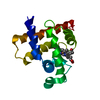
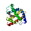
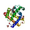
| Title | Sperm whale myoglobin mutant H64W deoxy-form | ||||||
 Components Components | Myoglobin | ||||||
 Keywords Keywords | OXYGEN TRANSPORT / Myoglobin / ligand migration pathways / oxygen storage | ||||||
| Function / homology |  Function and homology information Function and homology informationOxidoreductases; Acting on other nitrogenous compounds as donors / nitrite reductase activity / sarcoplasm / Oxidoreductases; Acting on a peroxide as acceptor; Peroxidases / removal of superoxide radicals / oxygen carrier activity / peroxidase activity / oxygen binding / heme binding / extracellular exosome / metal ion binding Similarity search - Function | ||||||
| Biological species |  | ||||||
| Method |  X-RAY DIFFRACTION / FOURIER SYNTHESIS / Resolution: 1.6 Å X-RAY DIFFRACTION / FOURIER SYNTHESIS / Resolution: 1.6 Å | ||||||
 Authors Authors | Birukou, I. / Soman, J. / Olson, J.S. | ||||||
 Citation Citation | Journal: J.Biol.Chem. / Year: 2011 Title: Blocking the gate to ligand entry in human hemoglobin. Authors: Birukou, I. / Soman, J. / Olson, J.S. | ||||||
| History |
|
- Structure visualization
| Structure viewer | Molecule: MolmilJmol/JSmol |
|---|
- Downloads & links
-Download
| PDBx/mmCIF format | 3ogb.cif.gz | 57.3 KB | Display | PDBx/mmCIF format |
|---|---|---|---|---|
| PDB format | pdb3ogb.ent.gz | 40.2 KB | Display | PDB format |
| PDBx/mmJSON format | 3ogb.json.gz | Tree view | PDBx/mmJSON format | |
| Others |  Other downloads Other downloads |
-Validation report
| Arichive directory | https://data.pdbj.org/pub/pdb/validation_reports/og/3ogbftp://data.pdbj.org/pub/pdb/validation_reports/og/3ogb | HTTPS FTP |
|---|
-Related structure data
| Related structure data |  3nl7C  3nmlSC  3nmmC C: citing same article ( S: Starting model for refinement |
|---|---|
| Similar structure data |
-Links
 PDBj
PDBj


- Assembly
| Deposited unit | 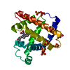
| ||||||||
|---|---|---|---|---|---|---|---|---|---|
| 1 |
| ||||||||
| Unit cell |
|
-Components
| #1: Protein | Mass: 17413.225 Da / Num. of mol.: 1 / Mutation: H65W Source method: isolated from a genetically manipulated source Source: (gene. exp.)  | ||
|---|---|---|---|
| #2: Chemical | ChemComp-HEM /   Mass: 616.487 Da / Num. of mol.: 1 / Source method: obtained synthetically / Formula: C34H32FeN4O4 Mass: 616.487 Da / Num. of mol.: 1 / Source method: obtained synthetically / Formula: C34H32FeN4O4 | ||
| #3: Chemical | ChemComp-SO4 /   Mass: 96.063 Da / Num. of mol.: 9 / Source method: obtained synthetically / Formula: SO4 Mass: 96.063 Da / Num. of mol.: 9 / Source method: obtained synthetically / Formula: SO4#4: Water | ChemComp-HOH / |  Mass: 18.015 Da / Num. of mol.: 249 / Source method: isolated from a natural source / Formula: H2O Mass: 18.015 Da / Num. of mol.: 249 / Source method: isolated from a natural source / Formula: H2O |
-Experimental details
-Experiment
| Experiment | Method: X-RAY DIFFRACTION / Number of used crystals: 1 |
|---|
- Sample preparation
| Crystal | Density Matthews: 2.34 Å3/Da / Density % sol: 47.36 % |
|---|---|
| Crystal grow | Temperature: 293 K / Method: small tubes / pH: 9 Details: Solution of MB (carbonmonoxy-form) (20mg/ml protein in 0.02M TRIS-HCl pH 9.0) mixed with mother liquor (3.2M ammonium sulphate, 0.05M TRIS-HCl, pH 9.0) for final concentrations of 2.5M ...Details: Solution of MB (carbonmonoxy-form) (20mg/ml protein in 0.02M TRIS-HCl pH 9.0) mixed with mother liquor (3.2M ammonium sulphate, 0.05M TRIS-HCl, pH 9.0) for final concentrations of 2.5M ammonium sulphate to obtain crystals of MBCO. MBCO crystals were oxidyzed by excess of potassium ferricyanide, washed in mother liquor and reduced under anaerobic conditions with sodium dithionite, SMALL TUBES, temperature 293K |
-Data collection
| Diffraction | Mean temperature: 100 K | ||||||||||||||||||||||||||||||||||||||||||||||||||||||||||||||||||
|---|---|---|---|---|---|---|---|---|---|---|---|---|---|---|---|---|---|---|---|---|---|---|---|---|---|---|---|---|---|---|---|---|---|---|---|---|---|---|---|---|---|---|---|---|---|---|---|---|---|---|---|---|---|---|---|---|---|---|---|---|---|---|---|---|---|---|---|
| Diffraction source | Source: ROTATING ANODE / Type: RIGAKU RUH3R / Wavelength: 1.5418 Å | ||||||||||||||||||||||||||||||||||||||||||||||||||||||||||||||||||
| Detector | Type: RIGAKU RAXIS IV++ / Detector: IMAGE PLATE / Date: Jul 23, 2010 | ||||||||||||||||||||||||||||||||||||||||||||||||||||||||||||||||||
| Radiation | Monochromator: GRAPHITE / Protocol: SINGLE WAVELENGTH / Monochromatic (M) / Laue (L): M / Scattering type: x-ray | ||||||||||||||||||||||||||||||||||||||||||||||||||||||||||||||||||
| Radiation wavelength | Wavelength: 1.5418 Å / Relative weight: 1 | ||||||||||||||||||||||||||||||||||||||||||||||||||||||||||||||||||
| Reflection | Resolution: 1.6→23.57 Å / Num. obs: 21466 / % possible obs: 96.6 % / Redundancy: 3.65 % / Rmerge(I) obs: 0.043 / Χ2: 0.95 / Net I/σ(I): 15.5 / Scaling rejects: 695 | ||||||||||||||||||||||||||||||||||||||||||||||||||||||||||||||||||
| Reflection shell |
|
- Processing
| Software |
| |||||||||||||||||||||||||||||||||||||||||||||||||||||||||||||||
|---|---|---|---|---|---|---|---|---|---|---|---|---|---|---|---|---|---|---|---|---|---|---|---|---|---|---|---|---|---|---|---|---|---|---|---|---|---|---|---|---|---|---|---|---|---|---|---|---|---|---|---|---|---|---|---|---|---|---|---|---|---|---|---|---|
| Refinement | Method to determine structure: FOURIER SYNTHESIS Starting model: PDB ENTRY 3NML Resolution: 1.6→23.565 Å / Occupancy max: 1 / Occupancy min: 0.2 / SU ML: 0.21 / Cross valid method: THROUGHOUT / σ(F): 0 / Phase error: 17.85 / Stereochemistry target values: ML
| |||||||||||||||||||||||||||||||||||||||||||||||||||||||||||||||
| Solvent computation | Shrinkage radii: 0.04 Å / VDW probe radii: 0.4 Å / Solvent model: FLAT BULK SOLVENT MODEL / Bsol: 77.122 Å2 / ksol: 0.435 e/Å3 | |||||||||||||||||||||||||||||||||||||||||||||||||||||||||||||||
| Displacement parameters | Biso max: 73.49 Å2 / Biso mean: 20.7404 Å2 / Biso min: 9.65 Å2
| |||||||||||||||||||||||||||||||||||||||||||||||||||||||||||||||
| Refinement step | Cycle: LAST / Resolution: 1.6→23.565 Å
| |||||||||||||||||||||||||||||||||||||||||||||||||||||||||||||||
| Refine LS restraints |
| |||||||||||||||||||||||||||||||||||||||||||||||||||||||||||||||
| LS refinement shell | Refine-ID: X-RAY DIFFRACTION / Total num. of bins used: 8
|
