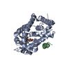
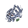
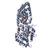
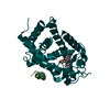
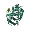
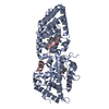
[English] 日本語
Yorodumi
- PDB-3o1d: Structure-function study of Gemini derivatives with two different... -

+
Open data


ID or keywords:

Loading...

-
Basic information

Entry
Database: PDB / ID: 3o1d
TitleStructure-function study of Gemini derivatives with two different side chains at C-20, Gemini-0072 and Gemini-0097.
Components
  • Nuclear receptor coactivator 2
  • Vitamin D3 receptor A
KeywordsTRANSCRIPTION/TRANSCRIPTION ACTIVATOR / Transcription factor / vitamin D / Nucleus / TRANSCRIPTION-TRANSCRIPTION ACTIVATOR complex
Function / homology
Function and homology information


heart jogging / Vitamin D (calciferol) metabolism / : / SUMOylation of intracellular receptors / vitamin D binding / calcitriol binding / lithocholic acid binding / hematopoietic stem cell proliferation / RNA polymerase II intronic transcription regulatory region sequence-specific DNA binding / heart looping ...heart jogging / Vitamin D (calciferol) metabolism / : / SUMOylation of intracellular receptors / vitamin D binding / calcitriol binding / lithocholic acid binding / hematopoietic stem cell proliferation / RNA polymerase II intronic transcription regulatory region sequence-specific DNA binding / heart looping / locomotor rhythm / aryl hydrocarbon receptor binding / regulation of lipid metabolic process / cellular response to Thyroglobulin triiodothyronine / regulation of glucose metabolic process / Synthesis of bile acids and bile salts / calcium ion homeostasis / Endogenous sterols / Synthesis of bile acids and bile salts via 27-hydroxycholesterol / Synthesis of bile acids and bile salts via 7alpha-hydroxycholesterol / Recycling of bile acids and salts / regulation of cellular response to insulin stimulus / cellular response to hormone stimulus / positive regulation of adipose tissue development / peroxisome proliferator activated receptor signaling pathway / RORA activates gene expression / Regulation of lipid metabolism by PPARalpha / ossification / BMAL1:CLOCK,NPAS2 activates circadian gene expression / SUMOylation of transcription cofactors / nuclear receptor coactivator activity / Activation of gene expression by SREBF (SREBP) / response to progesterone / nuclear receptor binding / circadian regulation of gene expression / Heme signaling / mRNA transcription by RNA polymerase II / Activated PKN1 stimulates transcription of AR (androgen receptor) regulated genes KLK2 and KLK3 / Transcriptional activation of mitochondrial biogenesis / PPARA activates gene expression / Cytoprotection by HMOX1 / Transcriptional regulation of white adipocyte differentiation / RNA polymerase II transcription regulator complex / nuclear receptor activity / Circadian Clock / HATs acetylate histones / Estrogen-dependent gene expression / transcription regulator complex / cell differentiation / transcription coactivator activity / protein dimerization activity / nuclear body / RNA polymerase II cis-regulatory region sequence-specific DNA binding / protein domain specific binding / DNA-templated transcription / chromatin binding / chromatin / regulation of DNA-templated transcription / negative regulation of transcription by RNA polymerase II / positive regulation of transcription by RNA polymerase II / protein-containing complex / zinc ion binding / nucleoplasm / nucleus / cytoplasm
Similarity search - Function
Vitamin D receptor / VDR, DNA-binding domain / Nuclear receptor coactivator 2 / Nuclear receptor coactivator 2/3, DUF4927 / Domain of unknown function (DUF4927) / Nuclear receptor coactivator, DUF1518 / Nuclear receptor coactivator, Ncoa-type, interlocking / Nuclear receptor coactivator, Ncoa-type, interlocking domain superfamily / Nuclear receptor coactivator, DUF1518 / Nuclear receptor coactivator ...Vitamin D receptor / VDR, DNA-binding domain / Nuclear receptor coactivator 2 / Nuclear receptor coactivator 2/3, DUF4927 / Domain of unknown function (DUF4927) / Nuclear receptor coactivator, DUF1518 / Nuclear receptor coactivator, Ncoa-type, interlocking / Nuclear receptor coactivator, Ncoa-type, interlocking domain superfamily / Nuclear receptor coactivator, DUF1518 / Nuclear receptor coactivator / DUF1518 / Nuclear receptor coactivator, receptor-binding domain / Nuclear receptor coactivator / Steroid receptor coactivator / Unstructured region on nuclear receptor coactivator protein / PAS domain / Nuclear receptor coactivator, interlocking / Helix-loop-helix DNA-binding domain superfamily / helix loop helix domain / Myc-type, basic helix-loop-helix (bHLH) domain / Myc-type, basic helix-loop-helix (bHLH) domain profile. / PAS fold / PAS fold / PAS domain / PAS repeat profile. / PAS domain / PAS domain superfamily / Retinoid X Receptor / Retinoid X Receptor / Nuclear hormone receptor / Nuclear hormones receptors DNA-binding region signature. / Zinc finger, nuclear hormone receptor-type / Zinc finger, C4 type (two domains) / Nuclear hormone receptors DNA-binding domain profile. / c4 zinc finger in nuclear hormone receptors / Nuclear hormone receptor, ligand-binding domain / Nuclear hormone receptor-like domain superfamily / Ligand-binding domain of nuclear hormone receptor / Nuclear receptor (NR) ligand-binding (LBD) domain profile. / Ligand binding domain of hormone receptors / Zinc finger, NHR/GATA-type / Orthogonal Bundle / Mainly Alpha
Similarity search - Domain/homology
Chem-G72 / Nuclear receptor coactivator 2 / Vitamin D3 receptor A
Similarity search - Component
Biological speciesDanio rerio (zebrafish)
Homo sapiens (human)
MethodX-RAY DIFFRACTION / SYNCHROTRON / FOURIER SYNTHESIS / Resolution: 2.4007 Å
AuthorsHuet, T. / Moras, D. / Rochel, N.
CitationJournal: Medchemcomm / Year: 2011
Title: Structure-function study of gemini derivatives with two different side chains at C-20, Gemini-0072 and Gemini-0097.
Authors: Huet, T. / Maehr, H. / Lee, H.J. / Uskokovic, M.R. / Suh, N. / Moras, D. / Rochel, N.
History
DepositionJul 21, 2010Deposition site: RCSB / Processing site: RCSB
Revision 1.0Jul 6, 2011Provider: repository / Type: Initial release
Revision 1.1Jul 13, 2011Group: Version format compliance
Revision 1.2Mar 21, 2012Group: Database references
Revision 1.3Mar 10, 2021Group: Advisory / Database references / Derived calculations
Category: pdbx_struct_assembly / pdbx_struct_assembly_gen ...pdbx_struct_assembly / pdbx_struct_assembly_gen / pdbx_struct_assembly_prop / pdbx_unobs_or_zero_occ_atoms / struct_ref_seq_dif / struct_site
Item: _struct_ref_seq_dif.details / _struct_site.pdbx_auth_asym_id ..._struct_ref_seq_dif.details / _struct_site.pdbx_auth_asym_id / _struct_site.pdbx_auth_comp_id / _struct_site.pdbx_auth_seq_id
Revision 1.4Feb 21, 2024Group: Advisory / Data collection ...Advisory / Data collection / Database references / Structure summary
Category: chem_comp / chem_comp_atom ...chem_comp / chem_comp_atom / chem_comp_bond / database_2 / pdbx_unobs_or_zero_occ_atoms
Item: _chem_comp.pdbx_synonyms / _database_2.pdbx_DOI / _database_2.pdbx_database_accession

-
Structure visualization

Structure viewerMolecule:
MolmilJmol/JSmol

Downloads & links

-
Assembly

Deposited unit
A: Vitamin D3 receptor A
B: Nuclear receptor coactivator 2
hetero molecules


Theoretical massNumber of molelcules
Total (without water)36,2353
Polymers35,6412
Non-polymers5951
Water1,13563
1
A: Vitamin D3 receptor A
B: Nuclear receptor coactivator 2
hetero molecules

A: Vitamin D3 receptor A
B: Nuclear receptor coactivator 2
hetero molecules


Theoretical massNumber of molelcules
Total (without water)72,4706
Polymers71,2814
Non-polymers1,1892
Water724
TypeNameSymmetry operationNumber
identity operation1_555x,y,z1
crystal symmetry operation10_665-y+1,-x+1,-z+1/61
Buried area3860 Å2
ΔGint-35 kcal/mol
Surface area22610 Å2
MethodPISA
Unit cell
Length a, b, c (Å)66.108, 66.108, 264.640
Angle α, β, γ (deg.)90.00, 90.00, 120.00
Int Tables number179
Space group name H-MP6522

-
Components

#1: Protein Vitamin D3 receptor A / VDR-A / 1 / 25-dihydroxyvitamin D3 receptor A / Nuclear receptor subfamily 1 group I member 1-A


Mass: 34060.672 Da / Num. of mol.: 1 / Fragment: Ligand Binding Domain (UNP Residues 156-453)
Source method: isolated from a genetically manipulated source
Source: (gene. exp.) Danio rerio (zebrafish) / Gene: vdra, nr1i1a, vdr / Production host: Escherichia coli (E. coli) / Strain (production host): BL21(DE3) / References: UniProt: Q9PTN2
#2: Protein/peptide Nuclear receptor coactivator 2 / / NCoA-2 / Transcriptional intermediary factor 2 / hTIF2 / Class E basic helix-loop-helix protein 75 / bHLHe75


Mass: 1579.866 Da / Num. of mol.: 1 / Source method: obtained synthetically / Details: 13-MER PEPTIDE / Source: (synth.) Homo sapiens (human) / References: UniProt: Q15596
#3: Chemical ChemComp-G72 / (1R,3R,7E,17beta)-17-[(1S)-6,6,6-trifluoro-5-hydroxy-1-(4-hydroxy-4-methylpentyl)-5-(trifluoromethyl)hex-3-yn-1-yl]-9,10-secoestra-5,7-diene-1,3-diol / 1,25-dihydroxy-20S-21(3-trideuteromethyl-3-hydroxy-4,4,4-trideuterobutyl)-23-yne-26,27-hexafluoro-19-nor-cholecalciferol / Gemini-0072


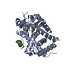
Mass: 594.669 Da / Num. of mol.: 1 / Source method: obtained synthetically / Formula: C31H44F6O4
#4: Water ChemComp-HOH / water / Water


Mass: 18.015 Da / Num. of mol.: 63 / Source method: isolated from a natural source / Formula: H2O

-
Experimental details

-
Experiment

ExperimentMethod: X-RAY DIFFRACTION / Number of used crystals: 1

-
Sample preparation

CrystalDensity Matthews: 2.34 Å3/Da / Density % sol: 47.48 %
Crystal growTemperature: 297 K / Method: vapor diffusion, hanging drop / pH: 6.5
Details: 50 mM BIS-TRIS pH6.5, 1.6 M lithium sulfate, 50 mM magnesium sulfate, VAPOR DIFFUSION, HANGING DROP, temperature 297K

-
Data collection

DiffractionMean temperature: 100 K
Diffraction sourceSource: SYNCHROTRON / Site: ESRF / Beamline: ID14-1 / Wavelength: 0.9334 Å
DetectorType: ADSC QUANTUM 210 / Detector: CCD / Date: Dec 11, 2008
RadiationMonochromator: Diamond / Protocol: SINGLE WAVELENGTH / Monochromatic (M) / Laue (L): M / Scattering type: x-ray
Radiation wavelengthWavelength: 0.9334 Å / Relative weight: 1
ReflectionResolution: 2.4→50 Å / Num. all: 14413 / Num. obs: 13853 / % possible obs: 96.2 % / Observed criterion σ(I): 14.8
Reflection shellResolution: 2.4→2.49 Å / % possible all: 99.6

-
Processing

Software
NameVersionClassification
MxCuBEdata collection
PHENIXmodel building
PHENIX(phenix.refine: 1.6.1_357)refinement
HKL-2000data reduction
HKL-2000data scaling
PHENIXphasing
RefinementMethod to determine structure: FOURIER SYNTHESIS / Resolution: 2.4007→24.884 Å / SU ML: 0.31 / σ(F): 1.34 / Phase error: 25.33 / Stereochemistry target values: ML
RfactorNum. reflection% reflectionSelection details
Rfree0.2671 699 5.08 %RANDOM
Rwork0.209 ---
obs0.2118 13747 96.16 %-
all-14413 --
Solvent computationShrinkage radii: 0.9 Å / VDW probe radii: 1.11 Å / Solvent model: FLAT BULK SOLVENT MODEL / Bsol: 73.384 Å2 / ksol: 0.371 e/Å3
Displacement parameters
Baniso -1Baniso -2Baniso -3
1-9.183 Å2-0 Å2-0 Å2
2--9.183 Å2-0 Å2
3----18.3661 Å2
Refinement stepCycle: LAST / Resolution: 2.4007→24.884 Å
ProteinNucleic acidLigandSolventTotal
Num. atoms2012 0 41 63 2116
Refine LS restraints
Refine-IDTypeDev idealNumber
X-RAY DIFFRACTIONf_bond_d0.0032099
X-RAY DIFFRACTIONf_angle_d0.8612840
X-RAY DIFFRACTIONf_dihedral_angle_d18.254815
X-RAY DIFFRACTIONf_chiral_restr0.05324
X-RAY DIFFRACTIONf_plane_restr0.002356
LS refinement shell
Resolution (Å)Rfactor RfreeNum. reflection RfreeRfactor RworkNum. reflection RworkRefine-ID% reflection obs (%)
2.4007-2.58590.34361580.28852569X-RAY DIFFRACTION99
2.5859-2.84580.31061620.26162611X-RAY DIFFRACTION99
2.8458-3.25680.28191190.2272680X-RAY DIFFRACTION100
3.2568-4.10030.28591190.19932497X-RAY DIFFRACTION91
4.1003-24.88550.22051410.17812691X-RAY DIFFRACTION92
Refinement TLS params.Method: refined / Origin x: 8.8392 Å / Origin y: 36.7782 Å / Origin z: 42.1761 Å
111213212223313233
T0.3494 Å20.1158 Å2-0.0604 Å2-0.4349 Å2-0.0483 Å2--0.3239 Å2
L0.8624 °2-0.0416 °20.3517 °2-2.0688 °21.6098 °2--2.829 °2
S-0.1703 Å °-0.3865 Å °-0.0078 Å °0.3948 Å °0.0823 Å °-0.0116 Å °0.2387 Å °-0.1112 Å °0.0547 Å °
Refinement TLS groupSelection details: all

+
About Yorodumi

-
News

-
Feb 9, 2022. New format data for meta-information of EMDB entries

New format data for meta-information of EMDB entries

  • Version 3 of the EMDB header file is now the official format.
  • The previous official version 1.9 will be removed from the archive.

Related info.:EMDB header

External links:wwPDB to switch to version 3 of the EMDB data model

-
Aug 12, 2020. Covid-19 info

Covid-19 info

URL: https://pdbj.org/emnavi/covid19.php

New page: Covid-19 featured information page in EM Navigator.

Related info.:Covid-19 info / Mar 5, 2020. Novel coronavirus structure data

+
Mar 5, 2020. Novel coronavirus structure data

Novel coronavirus structure data

Related info.:Yorodumi Speices / Aug 12, 2020. Covid-19 info

External links:COVID-19 featured content - PDBj / Molecule of the Month (242):Coronavirus Proteases

+
Jan 31, 2019. EMDB accession codes are about to change! (news from PDBe EMDB page)

EMDB accession codes are about to change! (news from PDBe EMDB page)

  • The allocation of 4 digits for EMDB accession codes will soon come to an end. Whilst these codes will remain in use, new EMDB accession codes will include an additional digit and will expand incrementally as the available range of codes is exhausted. The current 4-digit format prefixed with “EMD-” (i.e. EMD-XXXX) will advance to a 5-digit format (i.e. EMD-XXXXX), and so on. It is currently estimated that the 4-digit codes will be depleted around Spring 2019, at which point the 5-digit format will come into force.
  • The EM Navigator/Yorodumi systems omit the EMD- prefix.

Related info.:Q: What is EMD? / ID/Accession-code notation in Yorodumi/EM Navigator

External links:EMDB Accession Codes are Changing Soon! / Contact to PDBj

+
Jul 12, 2017. Major update of PDB

Major update of PDB

  • wwPDB released updated PDB data conforming to the new PDBx/mmCIF dictionary.
  • This is a major update changing the version number from 4 to 5, and with Remediation, in which all the entries are updated.
  • In this update, many items about electron microscopy experimental information are reorganized (e.g. em_software).
  • Now, EM Navigator and Yorodumi are based on the updated data.

External links:wwPDB Remediation / Enriched Model Files Conforming to OneDep Data Standards Now Available in the PDB FTP Archive

-
Yorodumi

Thousand views of thousand structures

  • Yorodumi is a browser for structure data from EMDB, PDB, SASBDB, etc.
  • This page is also the successor to EM Navigator detail page, and also detail information page/front-end page for Omokage search.
  • The word "yorodu" (or yorozu) is an old Japanese word meaning "ten thousand". "mi" (miru) is to see.

Related info.:EMDB / PDB / SASBDB / Comparison of 3 databanks / Yorodumi Search / Aug 31, 2016. New EM Navigator & Yorodumi / Yorodumi Papers / Jmol/JSmol / Function and homology information / Changes in new EM Navigator and Yorodumi

Read more