 Movie
Movie Controller

[English] 日本語
 Yorodumi
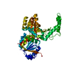
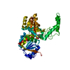
- PDB-3npi: Crystal structure of a TetR family regulatory protein (DIP1788) f... -
+ Open data

- Basic information
| Entry | Database: PDB / ID: 3npi | ||||||
|---|---|---|---|---|---|---|---|
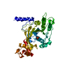
| Title | Crystal structure of a TetR family regulatory protein (DIP1788) from CORYNEBACTERIUM DIPHTHERIAE at 2.96 A resolution | ||||||
 Components Components | TetR family regulatory protein | ||||||
 Keywords Keywords | GENE REGULATION / Structural Genomics / Joint Center for Structural Genomics / JCSG / Protein Structure Initiative / PSI | ||||||
| Function / homology |  Function and homology information Function and homology information | ||||||
| Biological species |  Corynebacterium diphtheriae (bacteria) Corynebacterium diphtheriae (bacteria) | ||||||
| Method |  X-RAY DIFFRACTION / SYNCHROTRON / MAD / Resolution: 2.96 Å X-RAY DIFFRACTION / SYNCHROTRON / MAD / Resolution: 2.96 Å | ||||||
 Authors Authors | Joint Center for Structural Genomics (JCSG) | ||||||
 Citation Citation | Journal: To be published Title: Crystal structure of a TetR family regulatory protein (DIP1788) from CORYNEBACTERIUM DIPHTHERIAE at 2.96 A resolution Authors: Joint Center for Structural Genomics (JCSG) | ||||||
| History |
|
- Structure visualization
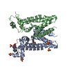
| Structure viewer | Molecule: MolmilJmol/JSmol |
|---|
- Downloads & links
-Download
| PDBx/mmCIF format | 3npi.cif.gz | 172.6 KB | Display | PDBx/mmCIF format |
|---|---|---|---|---|
| PDB format | pdb3npi.ent.gz | 142.6 KB | Display | PDB format |
| PDBx/mmJSON format | 3npi.json.gz | Tree view | PDBx/mmJSON format | |
| Others |  Other downloads Other downloads |
-Validation report
| Summary document | 3npi_validation.pdf.gz | 448.6 KB | Display | wwPDB validaton report |
|---|---|---|---|---|
| Full document | 3npi_full_validation.pdf.gz | 451.7 KB | Display | |
| Data in XML | 3npi_validation.xml.gz | 16.2 KB | Display | |
| Data in CIF | 3npi_validation.cif.gz | 20.8 KB | Display | |
| Arichive directory | https://data.pdbj.org/pub/pdb/validation_reports/np/3npiftp://data.pdbj.org/pub/pdb/validation_reports/np/3npi | HTTPS FTP |
-Related structure data
| Similar structure data | |
|---|---|
| Other databases |
-Links
 PDBj
PDBj- Assembly
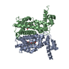
| Deposited unit | 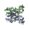
| ||||||||
|---|---|---|---|---|---|---|---|---|---|
| 1 |
| ||||||||
| Unit cell |
| ||||||||
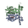
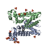
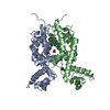
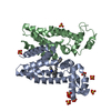
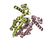
| Details | ANALYTICAL SIZE EXCLUSION CHROMATOGRAPHY WITH STATIC LIGHT SCATTERING SUPPORTS THE ASSIGNMENT OF A DIMER AS A SIGNIFICANT OLIGOMERIZATION STATE IN SOLUTION. |
-Components
| #1: Protein | Mass: 28217.109 Da / Num. of mol.: 2 Source method: isolated from a genetically manipulated source Source: (gene. exp.) Corynebacterium diphtheriae (bacteria) / Gene: DIP1788 / Plasmid: SpeedET / Production host: #2: Chemical | ChemComp-SO4 /   Mass: 96.063 Da / Num. of mol.: 8 / Source method: obtained synthetically / Formula: SO4 Mass: 96.063 Da / Num. of mol.: 8 / Source method: obtained synthetically / Formula: SO4Sequence details | THIS CONSTRUCT WAS EXPRESSED WITH A PURIFICATION TAG MGSDKIHHHHHHENLYFQG. THE TAG WAS REMOVED WITH ...THIS CONSTRUCT WAS EXPRESSED WITH A PURIFICATI | |
|---|
-Experimental details
-Experiment
| Experiment | Method: X-RAY DIFFRACTION / Number of used crystals: 1 |
|---|
- Sample preparation
| Crystal | Density Matthews: 2.64 Å3/Da / Density % sol: 53.34 % |
|---|---|
| Crystal grow | Temperature: 277 K / Method: vapor diffusion, sitting drop / pH: 6.86 Details: 1.7100M ammonium sulfate, 10.0000% Dioxane, 0.1M MES pH 6.86, VAPOR DIFFUSION,SITTING DROP,NANODROP, temperature 277K, VAPOR DIFFUSION, SITTING DROP |
-Data collection
| Diffraction | Mean temperature: 100 K | ||||||||||||||||||||||||||||||||||||||||||||||||||||||||||||||||||||||||||||||||||||||||||||||||||||||||||||||
|---|---|---|---|---|---|---|---|---|---|---|---|---|---|---|---|---|---|---|---|---|---|---|---|---|---|---|---|---|---|---|---|---|---|---|---|---|---|---|---|---|---|---|---|---|---|---|---|---|---|---|---|---|---|---|---|---|---|---|---|---|---|---|---|---|---|---|---|---|---|---|---|---|---|---|---|---|---|---|---|---|---|---|---|---|---|---|---|---|---|---|---|---|---|---|---|---|---|---|---|---|---|---|---|---|---|---|---|---|---|---|---|
| Diffraction source | Source: SYNCHROTRON / Site: SSRL  / Beamline: BL11-1 / Wavelength: 0.91837,0.97959,0.97925 / Beamline: BL11-1 / Wavelength: 0.91837,0.97959,0.97925 | ||||||||||||||||||||||||||||||||||||||||||||||||||||||||||||||||||||||||||||||||||||||||||||||||||||||||||||||
| Detector | Type: MARMOSAIC 325 mm CCD / Detector: CCD / Date: Feb 11, 2010 / Details: Flat mirror (vertical focusing) | ||||||||||||||||||||||||||||||||||||||||||||||||||||||||||||||||||||||||||||||||||||||||||||||||||||||||||||||
| Radiation | Monochromator: Single crystal Si(111) bent monochromator (horizontal focusing) Protocol: MAD / Monochromatic (M) / Laue (L): M / Scattering type: x-ray | ||||||||||||||||||||||||||||||||||||||||||||||||||||||||||||||||||||||||||||||||||||||||||||||||||||||||||||||
| Radiation wavelength |
| ||||||||||||||||||||||||||||||||||||||||||||||||||||||||||||||||||||||||||||||||||||||||||||||||||||||||||||||
| Reflection | Resolution: 2.96→29.713 Å / Num. obs: 12584 / % possible obs: 99.7 % / Observed criterion σ(I): -3 / Biso Wilson estimate: 99.731 Å2 / Rmerge F obs: 0.115 / Rmerge(I) obs: 0.056 / Rrim(I) all: 0.066 / Net I/σ(I): 17.06 / Num. measured all: 93470 | ||||||||||||||||||||||||||||||||||||||||||||||||||||||||||||||||||||||||||||||||||||||||||||||||||||||||||||||
| Reflection shell | Diffraction-ID: 1
|
-Phasing
| Phasing | Method: MAD |
|---|
- Processing
| Software |
| ||||||||||||||||||||||||||||||||||||||||||||||||||||||||||||||||||||||||||||||||||||||||||||||||||||||||||||
|---|---|---|---|---|---|---|---|---|---|---|---|---|---|---|---|---|---|---|---|---|---|---|---|---|---|---|---|---|---|---|---|---|---|---|---|---|---|---|---|---|---|---|---|---|---|---|---|---|---|---|---|---|---|---|---|---|---|---|---|---|---|---|---|---|---|---|---|---|---|---|---|---|---|---|---|---|---|---|---|---|---|---|---|---|---|---|---|---|---|---|---|---|---|---|---|---|---|---|---|---|---|---|---|---|---|---|---|---|---|
| Refinement | Method to determine structure: MAD / Resolution: 2.96→29.713 Å / Cor.coef. Fo:Fc: 0.9434 / Cor.coef. Fo:Fc free: 0.9233 / Occupancy max: 1 / Occupancy min: 0.5 / Cross valid method: THROUGHOUT / σ(F): 0 Details: 1.A MET-INHIBITION PROTOCOL WAS USED FOR SELENOMETHIONINE INCORPORATION DURING PROTEIN EXPRESSION. THE OCCUPANCY OF THE SE ATOMS IN THE MSE RESIDUES WAS REDUCED TO 0.75 FOR THE REDUCED ...Details: 1.A MET-INHIBITION PROTOCOL WAS USED FOR SELENOMETHIONINE INCORPORATION DURING PROTEIN EXPRESSION. THE OCCUPANCY OF THE SE ATOMS IN THE MSE RESIDUES WAS REDUCED TO 0.75 FOR THE REDUCED SCATTERING POWER DUE TO PARTIAL S-MET INCORPORATION. 2. SULFATES FROM THE CRYSTALLIZATION WERE MODELED INTO THE STRUCTURE. 3.ATOM RECORD CONTAINS SUM OF TLS AND RESIDUAL B FACTORS. ANISOU RECORD CONTAINS SUM OF TLS AND RESIDUAL U FACTORS.
| ||||||||||||||||||||||||||||||||||||||||||||||||||||||||||||||||||||||||||||||||||||||||||||||||||||||||||||
| Displacement parameters | Biso max: 204.84 Å2 / Biso mean: 97.8883 Å2 / Biso min: 45.22 Å2
| ||||||||||||||||||||||||||||||||||||||||||||||||||||||||||||||||||||||||||||||||||||||||||||||||||||||||||||
| Refinement step | Cycle: LAST / Resolution: 2.96→29.713 Å
| ||||||||||||||||||||||||||||||||||||||||||||||||||||||||||||||||||||||||||||||||||||||||||||||||||||||||||||
| Refine LS restraints |
| ||||||||||||||||||||||||||||||||||||||||||||||||||||||||||||||||||||||||||||||||||||||||||||||||||||||||||||
| LS refinement shell | Resolution: 2.96→3.24 Å / Total num. of bins used: 6
| ||||||||||||||||||||||||||||||||||||||||||||||||||||||||||||||||||||||||||||||||||||||||||||||||||||||||||||
| Refinement TLS params. | Method: refined / Refine-ID: X-RAY DIFFRACTION
| ||||||||||||||||||||||||||||||||||||||||||||||||||||||||||||||||||||||||||||||||||||||||||||||||||||||||||||
| Refinement TLS group |
|
