 Movie
Movie Controller

[English] 日本語
 Yorodumi
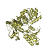
- PDB-3mcj: Crystal structure of molybdenum cofactor biosynthesis (AQ_061) ot... -
+ Open data

- Basic information
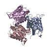
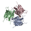
| Entry | Database: PDB / ID: 3mcj | ||||||
|---|---|---|---|---|---|---|---|
| Title | Crystal structure of molybdenum cofactor biosynthesis (AQ_061) other form from aquifex aeolicus VF5 | ||||||
 Components Components | Molybdenum cofactor biosynthesis MOG | ||||||
 Keywords Keywords | LYASE / Molybdopterin / MPT / Structural genomics / NPPSFA / National project on protein structural and functional analyses / RIKEN Structural genomics/proteomics initiative / RSGI | ||||||
| Function / homology |  Function and homology information Function and homology information | ||||||
| Biological species |   Aquifex aeolicus (bacteria) Aquifex aeolicus (bacteria) | ||||||
| Method |  X-RAY DIFFRACTION / SYNCHROTRON / MOLECULAR REPLACEMENT / Resolution: 1.9 Å X-RAY DIFFRACTION / SYNCHROTRON / MOLECULAR REPLACEMENT / Resolution: 1.9 Å | ||||||
 Authors Authors | Jeyakanthan, J. / Kanaujia, S.P. / Sekar, K. / Agari, Y. / Ebihara, A. / Kuramitsu, S. / Shinkai, A. / Yokoyama, S. / RIKEN Structural Genomics/Proteomics Initiative (RSGI) | ||||||
 Citation Citation | Journal: Acta Crystallogr.,Sect.F / Year: 2011 Title: Crystal structures, dynamics and functional implications of molybdenum-cofactor biosynthesis protein MogA from two thermophilic organisms Authors: Kanaujia, S.P. / Jeyakanthan, J. / Shinkai, A. / Kuramitsu, S. / Yokoyama, S. / Sekar, K. | ||||||
| History |
|
- Structure visualization
| Structure viewer | Molecule: MolmilJmol/JSmol |
|---|
- Downloads & links
-Download
| PDBx/mmCIF format | 3mcj.cif.gz | 221.4 KB | Display | PDBx/mmCIF format |
|---|---|---|---|---|
| PDB format | pdb3mcj.ent.gz | 176.9 KB | Display | PDB format |
| PDBx/mmJSON format | 3mcj.json.gz | Tree view | PDBx/mmJSON format | |
| Others |  Other downloads Other downloads |
-Validation report
| Summary document | 3mcj_validation.pdf.gz | 482.9 KB | Display | wwPDB validaton report |
|---|---|---|---|---|
| Full document | 3mcj_full_validation.pdf.gz | 498.7 KB | Display | |
| Data in XML | 3mcj_validation.xml.gz | 49.4 KB | Display | |
| Data in CIF | 3mcj_validation.cif.gz | 71.6 KB | Display | |
| Arichive directory | https://data.pdbj.org/pub/pdb/validation_reports/mc/3mcjftp://data.pdbj.org/pub/pdb/validation_reports/mc/3mcj | HTTPS FTP |
-Related structure data
| Related structure data | 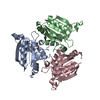 3mchC 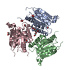 3mciSC S: Starting model for refinement C: citing same article ( |
|---|---|
| Similar structure data |


-Links
 PDBj
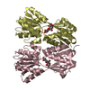
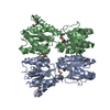
PDBj- Assembly
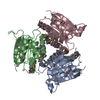
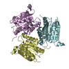
| Deposited unit | 
| ||||||||
|---|---|---|---|---|---|---|---|---|---|
| 1 | 
| ||||||||
| 2 | 
| ||||||||
| Unit cell |
|
-Components
| #1: Protein | Mass: 19298.580 Da / Num. of mol.: 6 Source method: isolated from a genetically manipulated source Source: (gene. exp.) Aquifex aeolicus (bacteria) / Strain: VF5 / Gene: mog, aq_061 / Plasmid: PET21A / Production host: #2: Chemical | ChemComp-EDO / |   Mass: 62.068 Da / Num. of mol.: 1 / Source method: obtained synthetically / Formula: C2H6O2 Mass: 62.068 Da / Num. of mol.: 1 / Source method: obtained synthetically / Formula: C2H6O2#3: Water | ChemComp-HOH / |  Mass: 18.015 Da / Num. of mol.: 953 / Source method: isolated from a natural source / Formula: H2O Mass: 18.015 Da / Num. of mol.: 953 / Source method: isolated from a natural source / Formula: H2O |
|---|
-Experimental details
-Experiment
| Experiment | Method: X-RAY DIFFRACTION / Number of used crystals: 1 |
|---|
- Sample preparation
| Crystal | Density Matthews: 2.13 Å3/Da / Density % sol: 42.17 % |
|---|---|
| Crystal grow | Temperature: 293 K / Method: vapor diffusion, sitting drop / pH: 5.5 Details: 0.2M AMMONIUM ACETATE, 0.1M BIS-TRIS, 25% PEG3350, pH 5.5, VAPOR DIFFUSION, SITTING DROP, temperature 293K |
-Data collection
| Diffraction | Mean temperature: 100 K |
|---|---|
| Diffraction source | Source: SYNCHROTRON / Site: SPring-8  / Beamline: BL26B2 / Wavelength: 1 Å / Beamline: BL26B2 / Wavelength: 1 Å |
| Detector | Type: RIGAKU JUPITER 210 / Detector: CCD / Date: Dec 19, 2006 / Details: RH COATED BENT-CYRINDRICAL MIRROR |
| Radiation | Protocol: SINGLE WAVELENGTH / Monochromatic (M) / Laue (L): M / Scattering type: x-ray |
| Radiation wavelength | Wavelength: 1 Å / Relative weight: 1 |
| Reflection | Resolution: 1.9→50 Å / Num. obs: 71606 / % possible obs: 96.4 % / Biso Wilson estimate: 12.6 Å2 / Rmerge(I) obs: 0.044 / Rsym value: 0.043 |
| Reflection shell | Resolution: 1.9→1.97 Å / Rmerge(I) obs: 0.219 / Rsym value: 0.199 / % possible all: 95.5 |
- Processing
| Software |
| ||||||||||||||||||||
|---|---|---|---|---|---|---|---|---|---|---|---|---|---|---|---|---|---|---|---|---|---|
| Refinement | Method to determine structure: MOLECULAR REPLACEMENT Starting model: 3MCI Resolution: 1.9→41.59 Å / Rfactor Rfree error: 0.004 / Data cutoff high absF: 189662.98 / Data cutoff low absF: 0 / Isotropic thermal model: RESTRAINED / Cross valid method: THROUGHOUT / σ(F): 0 / Stereochemistry target values: Engh & Huber / Details: BULK SOLVENT MODEL USED
| ||||||||||||||||||||
| Solvent computation | Solvent model: FLAT MODEL / Bsol: 58.0094 Å2 / ksol: 0.35 e/Å3 | ||||||||||||||||||||
| Displacement parameters | Biso mean: 32 Å2
| ||||||||||||||||||||
| Refine analyze |
| ||||||||||||||||||||
| Refinement step | Cycle: LAST / Resolution: 1.9→41.59 Å
| ||||||||||||||||||||
| Refine LS restraints |
| ||||||||||||||||||||
| LS refinement shell | Resolution: 1.9→2.02 Å / Rfactor Rfree error: 0.013 / Total num. of bins used: 6
| ||||||||||||||||||||
| Xplor file |
|
