 Movie
Movie Controller

[English] 日本語
 Yorodumi
- PDB-3ju8: Crystal Structure of Succinylglutamic Semialdehyde Dehydrogenase ... -
+ Open data

- Basic information
| Entry | Database: PDB / ID: 3ju8 | ||||||
|---|---|---|---|---|---|---|---|
| Title | Crystal Structure of Succinylglutamic Semialdehyde Dehydrogenase from Pseudomonas aeruginosa. | ||||||
 Components Components | Succinylglutamic semialdehyde dehydrogenase | ||||||
 Keywords Keywords |  OXIDOREDUCTASE / alpha-beta structure / Structural Genomics / PSI-2 / Protein Structure Initiative / Midwest Center for Structural Genomics / MCSG / Arginine metabolism / NAD OXIDOREDUCTASE / alpha-beta structure / Structural Genomics / PSI-2 / Protein Structure Initiative / Midwest Center for Structural Genomics / MCSG / Arginine metabolism / NAD | ||||||
| Function / homology |  Function and homology informationsuccinylglutamate-semialdehyde dehydrogenase / succinylglutamate-semialdehyde dehydrogenase activity / arginine catabolic process to succinate / arginine catabolic process to glutamate / arginine catabolic process Function and homology informationsuccinylglutamate-semialdehyde dehydrogenase / succinylglutamate-semialdehyde dehydrogenase activity / arginine catabolic process to succinate / arginine catabolic process to glutamate / arginine catabolic processSimilarity search - Function | ||||||
| Biological species |   Pseudomonas aeruginosa (bacteria) Pseudomonas aeruginosa (bacteria) | ||||||
| Method | X-RAY DIFFRACTION / SYNCHROTRON / SAD / Resolution: 1.82 Å | ||||||
 Authors Authors | Kim, Y. / Li, H. / Buck, K. / Joachimiak, A. / Midwest Center for Structural Genomics (MCSG) | ||||||
 Citation Citation | Journal: To be Published Title: Crystal Structure of Succinylglutamic Semialdehyde Dehydrogenase from Pseudomonas aeruginosa Authors: Kim, Y. / Li, H. / Buck, K. / Joachimiak, A. | ||||||
| History |
|
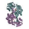
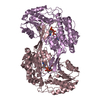
- Structure visualization
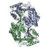
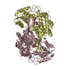
| Structure viewer | Molecule: MolmilJmol/JSmol |
|---|
- Downloads & links
-Download
| PDBx/mmCIF format | 3ju8.cif.gz | 412.4 KB | Display | PDBx/mmCIF format |
|---|---|---|---|---|
| PDB format | pdb3ju8.ent.gz | 350.5 KB | Display | PDB format |
| PDBx/mmJSON format | 3ju8.json.gz | Tree view | PDBx/mmJSON format | |
| Others |  Other downloads Other downloads |
-Validation report
| Arichive directory | https://data.pdbj.org/pub/pdb/validation_reports/ju/3ju8ftp://data.pdbj.org/pub/pdb/validation_reports/ju/3ju8 | HTTPS FTP |
|---|
-Related structure data
| Similar structure data | |
|---|---|
| Other databases |


-Links
 PDBj
PDBj

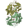
- Assembly
| Deposited unit | 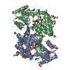
| ||||||||
|---|---|---|---|---|---|---|---|---|---|
| 1 |
| ||||||||
| Unit cell |
| ||||||||
| Components on special symmetry positions |
|
-Components
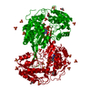
-Protein , 1 types, 2 molecules AB
| #1: Protein | Mass: 51999.973 Da / Num. of mol.: 2 Source method: isolated from a genetically manipulated source Source: (gene. exp.) Pseudomonas aeruginosa (bacteria) / Strain: PA01 / Gene: aruD, astD, PA0898 / Plasmid: pMCSG9 / Production host: Escherichia coli (E. coli) / Strain (production host): BL21 magicReferences: UniProt: O50174, succinylglutamate-semialdehyde dehydrogenase |
|---|
-Non-polymers , 7 types, 951 molecules 


| #2: Chemical | Nicotinamide adenine dinucleotide Mass: 663.425 Da / Num. of mol.: 2 / Source method: obtained synthetically / Formula: C21H27N7O14P2 / Comment: NAD*YM Mass: 663.425 Da / Num. of mol.: 2 / Source method: obtained synthetically / Formula: C21H27N7O14P2 / Comment: NAD*YM#3: Chemical | ChemComp-SO4 / Sulfate Mass: 96.063 Da / Num. of mol.: 8 / Source method: obtained synthetically / Formula: SO4 Mass: 96.063 Da / Num. of mol.: 8 / Source method: obtained synthetically / Formula: SO4#4: Chemical | ChemComp-SIN / | Succinic acid Mass: 118.088 Da / Num. of mol.: 1 / Source method: obtained synthetically / Formula: C4H6O4 Mass: 118.088 Da / Num. of mol.: 1 / Source method: obtained synthetically / Formula: C4H6O4#5: Chemical | ChemComp-GOL / Glycerol Mass: 92.094 Da / Num. of mol.: 8 / Source method: obtained synthetically / Formula: C3H8O3 Mass: 92.094 Da / Num. of mol.: 8 / Source method: obtained synthetically / Formula: C3H8O3#6: Chemical | Chloride Mass: 35.453 Da / Num. of mol.: 2 / Source method: obtained synthetically / Formula: Cl Mass: 35.453 Da / Num. of mol.: 2 / Source method: obtained synthetically / Formula: Cl#7: Chemical | ChemComp-MG / |  Mass: 24.305 Da / Num. of mol.: 1 / Source method: obtained synthetically / Formula: Mg Mass: 24.305 Da / Num. of mol.: 1 / Source method: obtained synthetically / Formula: Mg#8: Water | ChemComp-HOH / | WaterMass: 18.015 Da / Num. of mol.: 929 / Source method: isolated from a natural source / Formula: H2O |
|---|
-Experimental details
-Experiment
| Experiment | Method: X-RAY DIFFRACTION / Number of used crystals: 1 |
|---|
- Sample preparation
| Crystal | Density Matthews: 2.83 Å3/Da / Density % sol: 56.48 % |
|---|---|
| Crystal grow | Temperature: 289 K / Method: vapor diffusion, sitting drop / pH: 6.5 Details: 1.6 M Magnesium sulfate 0.1 M MES pH 6.5, VAPOR DIFFUSION, SITTING DROP, temperature 289K |
-Data collection
| Diffraction | Mean temperature: 100 K |
|---|---|
| Diffraction source | Source: SYNCHROTRON / Site: APS  / Beamline: 19-BM / Wavelength: 0.9788 Å / Beamline: 19-BM / Wavelength: 0.9788 Å |
| Detector | Type: ADSC QUANTUM 210r / Detector: CCD / Date: Aug 19, 2009 / Details: mirrors |
| Radiation | Monochromator: double crystal monochromator / Protocol: SINGLE WAVELENGTH / Monochromatic (M) / Laue (L): M / Scattering type: x-ray |
| Radiation wavelength | Wavelength: 0.9788 Å / Relative weight: 1 |
| Reflection | Resolution: 1.82→32.78 Å / Num. all: 104456 / Num. obs: 104456 / % possible obs: 99.3 % / Observed criterion σ(F): 0 / Observed criterion σ(I): 0 / Redundancy: 4 % / Biso Wilson estimate: 24.03 Å2 / Rsym value: 0.064 / Net I/σ(I): 12.5 |
| Reflection shell | Resolution: 1.82→1.85 Å / Redundancy: 3.2 % / Mean I/σ(I) obs: 2.9 / Num. unique all: 4769 / Rsym value: 0.41 / % possible all: 91.9 |
- Processing
| Software |
| ||||||||||||||||||||||||||||||||||||||||||||||||||||||||||||||||||||||||||||||
|---|---|---|---|---|---|---|---|---|---|---|---|---|---|---|---|---|---|---|---|---|---|---|---|---|---|---|---|---|---|---|---|---|---|---|---|---|---|---|---|---|---|---|---|---|---|---|---|---|---|---|---|---|---|---|---|---|---|---|---|---|---|---|---|---|---|---|---|---|---|---|---|---|---|---|---|---|---|---|---|
| Refinement | Method to determine structure: SAD / Resolution: 1.82→32.775 Å / SU ML: 0.21 / Cross valid method: THROUGHOUT / σ(F): 0 / σ(I): 0 / Phase error: 20.38 / Stereochemistry target values: MLHL
| ||||||||||||||||||||||||||||||||||||||||||||||||||||||||||||||||||||||||||||||
| Solvent computation | Shrinkage radii: 0.9 Å / VDW probe radii: 1.11 Å / Solvent model: FLAT BULK SOLVENT MODEL / Bsol: 59.845 Å2 / ksol: 0.383 e/Å3 | ||||||||||||||||||||||||||||||||||||||||||||||||||||||||||||||||||||||||||||||
| Displacement parameters |
| ||||||||||||||||||||||||||||||||||||||||||||||||||||||||||||||||||||||||||||||
| Refinement step | Cycle: LAST / Resolution: 1.82→32.775 Å
| ||||||||||||||||||||||||||||||||||||||||||||||||||||||||||||||||||||||||||||||
| Refine LS restraints |
| ||||||||||||||||||||||||||||||||||||||||||||||||||||||||||||||||||||||||||||||
| LS refinement shell | Refine-ID: X-RAY DIFFRACTION
| ||||||||||||||||||||||||||||||||||||||||||||||||||||||||||||||||||||||||||||||
| Refinement TLS params. | Refine-ID: X-RAY DIFFRACTION
| ||||||||||||||||||||||||||||||||||||||||||||||||||||||||||||||||||||||||||||||
| Refinement TLS group | Selection details: chain B |
