 Movie
Movie Controller

[English] 日本語
 Yorodumi
- PDB-3jcn: Structures of ribosome-bound initiation factor 2 reveal the mecha... -
+ Open data

- Basic information
| Entry | Database: PDB / ID: 3jcn | ||||||
|---|---|---|---|---|---|---|---|
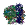
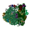
| Title | Structures of ribosome-bound initiation factor 2 reveal the mechanism of subunit association: Initiation Complex I | ||||||
 Components Components |
| ||||||
 Keywords Keywords | RIBOSOME / translation initiation / GTPase / 70S / bacterial ribosome / IF2 / initiation factor 2 | ||||||
| Function / homology |  Function and homology information Function and homology informationguanosine tetraphosphate binding / stringent response / ornithine decarboxylase inhibitor activity / transcription antitermination factor activity, RNA binding / misfolded RNA binding / Group I intron splicing / RNA folding / ribosomal small subunit binding / transcriptional attenuation / endoribonuclease inhibitor activity ...guanosine tetraphosphate binding / stringent response / ornithine decarboxylase inhibitor activity / transcription antitermination factor activity, RNA binding / misfolded RNA binding / Group I intron splicing / RNA folding / ribosomal small subunit binding / transcriptional attenuation / endoribonuclease inhibitor activity / RNA-binding transcription regulator activity / positive regulation of ribosome biogenesis / negative regulation of cytoplasmic translation / four-way junction DNA binding / translational termination / DnaA-L2 complex / chaperone-mediated protein folding / translation repressor activity / negative regulation of translational initiation / negative regulation of DNA-templated DNA replication initiation / regulation of mRNA stability / translation initiation factor activity / response to cold / mRNA regulatory element binding translation repressor activity / ribosome assembly / positive regulation of RNA splicing / assembly of large subunit precursor of preribosome / transcription elongation factor complex / cytosolic ribosome assembly / regulation of DNA-templated transcription elongation / DNA endonuclease activity / ribosomal large subunit assembly / transcription antitermination / response to reactive oxygen species / translational initiation / regulation of cell growth / DNA-templated transcription termination / maintenance of translational fidelity / response to radiation / mRNA 5'-UTR binding / large ribosomal subunit / ribosome biogenesis / ribosome binding / regulation of translation / ribosomal small subunit biogenesis / ribosomal small subunit assembly / small ribosomal subunit / small ribosomal subunit rRNA binding / transferase activity / 5S rRNA binding / large ribosomal subunit rRNA binding / cytosolic small ribosomal subunit / cytosolic large ribosomal subunit / cytoplasmic translation / tRNA binding / molecular adaptor activity / rRNA binding / negative regulation of translation / ribosome / structural constituent of ribosome / translation / response to antibiotic / negative regulation of DNA-templated transcription / GTPase activity / mRNA binding / GTP binding / DNA binding / RNA binding / zinc ion binding / membrane / cytoplasm / cytosol Similarity search - Function | ||||||
| Biological species |  | ||||||
| Method | ELECTRON MICROSCOPY / single particle reconstruction / cryo EM / Resolution: 4.6 Å | ||||||
 Authors Authors | Sprink, T. / Ramrath, D.J.F. / Yamamoto, H. / Yamamoto, K. / Loerke, J. / Ismer, J. / Hildebrand, P.W. / Scheerer, P. / Buerger, J. / Mielke, T. / Spahn, C.M.T. | ||||||
 Citation Citation | Journal: Sci Adv / Year: 2016 Title: Structures of ribosome-bound initiation factor 2 reveal the mechanism of subunit association. Authors: Thiemo Sprink / David J F Ramrath / Hiroshi Yamamoto / Kaori Yamamoto / Justus Loerke / Jochen Ismer / Peter W Hildebrand / Patrick Scheerer / Jörg Bürger / Thorsten Mielke / Christian M T Spahn /  Abstract: Throughout the four phases of protein biosynthesis-initiation, elongation, termination, and recycling-the ribosome is controlled and regulated by at least one specified translational guanosine ...Throughout the four phases of protein biosynthesis-initiation, elongation, termination, and recycling-the ribosome is controlled and regulated by at least one specified translational guanosine triphosphatase (trGTPase). Although the structural basis for trGTPase interaction with the ribosome has been solved for the last three steps of translation, the high-resolution structure for the key initiation trGTPase, initiation factor 2 (IF2), complexed with the ribosome, remains elusive. We determine the structure of IF2 complexed with a nonhydrolyzable guanosine triphosphate analog and initiator fMet-tRNAi (Met) in the context of the Escherichia coli ribosome to 3.7-Å resolution using cryo-electron microscopy. The structural analysis reveals previously unseen intrinsic conformational modes of the 70S initiation complex, establishing the mutual interplay of IF2 and initator transfer RNA (tRNA) with the ribsosome and providing the structural foundation for a mechanistic understanding of the final steps of translation initiation. | ||||||
| History |
|
- Structure visualization
| Movie |
Movie viewer |
|---|---|
| Structure viewer | Molecule: MolmilJmol/JSmol |
 UCSF Chimera
- Downloads & links
-Download
| PDBx/mmCIF format | 3jcn.cif.gz | 3.2 MB | Display | PDBx/mmCIF format |
|---|---|---|---|---|
| PDB format | pdb3jcn.ent.gz | Display | PDB format | |
| PDBx/mmJSON format | 3jcn.json.gz | Tree view | PDBx/mmJSON format | |
| Others |  Other downloads Other downloads |
-Validation report
| Summary document | 3jcn_validation.pdf.gz | 1.4 MB | Display | wwPDB validaton report |
|---|---|---|---|---|
| Full document | 3jcn_full_validation.pdf.gz | 1.4 MB | Display | |
| Data in XML | 3jcn_validation.xml.gz | 208.8 KB | Display | |
| Data in CIF | 3jcn_validation.cif.gz | 363.9 KB | Display | |
| Arichive directory | https://data.pdbj.org/pub/pdb/validation_reports/jc/3jcnftp://data.pdbj.org/pub/pdb/validation_reports/jc/3jcn | HTTPS FTP |
-Related structure data
| Related structure data | 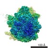 3285MC 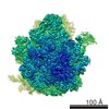 6559C 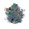 3jcjC M: map data used to model this data C: citing same article ( |
|---|---|
| Similar structure data |
-Links
 PDBj
PDBj


- Assembly
| Deposited unit | 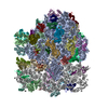
|
|---|---|
| 1 |
|
-Components
+50S ribosomal protein ... , 29 types, 29 molecules 01234CDEFGHIJKLMNOPQRSTUVWXYZ
-RNA chain , 5 types, 5 molecules ABavz
| #6: RNA chain | Mass: 941612.375 Da / Num. of mol.: 1 / Source method: isolated from a natural source / Source: (natural) |
|---|---|
| #7: RNA chain | Mass: 38790.090 Da / Num. of mol.: 1 / Source method: isolated from a natural source / Source: (natural) |
| #32: RNA chain | Mass: 499690.031 Da / Num. of mol.: 1 / Source method: isolated from a natural source / Source: (natural) |
| #52: RNA chain | Mass: 24818.893 Da / Num. of mol.: 1 / Source method: isolated from a natural source / Source: (natural) |
| #55: RNA chain | Mass: 1900.198 Da / Num. of mol.: 1 / Source method: isolated from a natural source / Source: (natural) |
-Protein , 1 types, 1 molecules b
| #33: Protein | Mass: 97498.164 Da / Num. of mol.: 1 Source method: isolated from a genetically manipulated source Source: (gene. exp.) |
|---|
-30S ribosomal protein ... , 20 types, 20 molecules cdfghijklmnopqrstuwx
| #34: Protein | Mass: 26031.316 Da / Num. of mol.: 1 / Source method: isolated from a natural source / Source: (natural) |
|---|---|
| #35: Protein | Mass: 26781.670 Da / Num. of mol.: 1 / Source method: isolated from a natural source / Source: (natural) |
| #36: Protein | Mass: 16772.412 Da / Num. of mol.: 1 / Source method: isolated from a natural source / Source: (natural) |
| #37: Protein | Mass: 23514.199 Da / Num. of mol.: 1 / Source method: isolated from a natural source / Source: (natural) |
| #38: Protein | Mass: 20055.156 Da / Num. of mol.: 1 / Source method: isolated from a natural source / Source: (natural) |
| #39: Protein | Mass: 15727.512 Da / Num. of mol.: 1 / Source method: isolated from a natural source / Source: (natural) |
| #40: Protein | Mass: 14886.270 Da / Num. of mol.: 1 / Source method: isolated from a natural source / Source: (natural) |
| #41: Protein | Mass: 14146.557 Da / Num. of mol.: 1 / Source method: isolated from a natural source / Source: (natural) |
| #42: Protein | Mass: 13870.975 Da / Num. of mol.: 1 / Source method: isolated from a natural source / Source: (natural) |
| #43: Protein | Mass: 11698.545 Da / Num. of mol.: 1 / Source method: isolated from a natural source / Source: (natural) |
| #44: Protein | Mass: 13128.467 Da / Num. of mol.: 1 / Source method: isolated from a natural source / Source: (natural) |
| #45: Protein | Mass: 13768.157 Da / Num. of mol.: 1 / Source method: isolated from a natural source / Source: (natural) |
| #46: Protein | Mass: 10290.816 Da / Num. of mol.: 1 / Source method: isolated from a natural source / Source: (natural) |
| #47: Protein | Mass: 11606.560 Da / Num. of mol.: 1 / Source method: isolated from a natural source / Source: (natural) |
| #48: Protein | Mass: 9724.491 Da / Num. of mol.: 1 / Source method: isolated from a natural source / Source: (natural) |
| #49: Protein | Mass: 9207.572 Da / Num. of mol.: 1 / Source method: isolated from a natural source / Source: (natural) |
| #50: Protein | Mass: 10455.355 Da / Num. of mol.: 1 / Source method: isolated from a natural source / Source: (natural) |
| #51: Protein | Mass: 9005.472 Da / Num. of mol.: 1 / Source method: isolated from a natural source / Source: (natural) |
| #53: Protein | Mass: 8524.039 Da / Num. of mol.: 1 / Source method: isolated from a natural source / Source: (natural) |
| #54: Protein | Mass: 9708.464 Da / Num. of mol.: 1 / Source method: isolated from a natural source / Source: (natural) |
-Non-polymers , 2 types, 2 molecules 
| #56: Chemical | ChemComp-GNP /  Mass: 522.196 Da / Num. of mol.: 1 / Source method: obtained synthetically / Formula: C10H17N6O13P3 Mass: 522.196 Da / Num. of mol.: 1 / Source method: obtained synthetically / Formula: C10H17N6O13P3Comment: GppNHp, GMPPNP, energy-carrying molecule analogue*YM |
|---|---|
| #57: Chemical | ChemComp-FME /  Type: L-peptide linking / Mass: 177.221 Da / Num. of mol.: 1 / Source method: obtained synthetically / Formula: C6H11NO3S Type: L-peptide linking / Mass: 177.221 Da / Num. of mol.: 1 / Source method: obtained synthetically / Formula: C6H11NO3S |
-Experimental details
-Experiment
| Experiment | Method: ELECTRON MICROSCOPY |
|---|---|
| EM experiment | Aggregation state: PARTICLE / 3D reconstruction method: single particle reconstruction |
- Sample preparation
| Component |
| |||||||||||||||||||||||||
|---|---|---|---|---|---|---|---|---|---|---|---|---|---|---|---|---|---|---|---|---|---|---|---|---|---|---|
| Buffer solution | Name: 20 mM HEPES-KOH, pH 7.5, 15 mM magnesium acetate, 150 mM potassium acetate, 4 mM 2-mercapthoethanol, 2 mM spermidine, 0.05 mM spermine pH: 7.5 Details: 20 mM HEPES-KOH, pH 7.5, 15 mM magnesium acetate, 150 mM potassium acetate, 4 mM 2-mercapthoethanol, 2 mM spermidine, 0.05 mM spermine | |||||||||||||||||||||||||
| Specimen | Embedding applied: NO / Shadowing applied: NO / Staining applied: NO / Vitrification applied: YES | |||||||||||||||||||||||||
| Specimen support | Details: Quantifoil R3-3 Cu 300 mesh with 2 nm carbon support film | |||||||||||||||||||||||||
| Vitrification | Instrument: FEI VITROBOT MARK I / Cryogen name: ETHANE / Humidity: 100 % Details: Blot for 2-4 seconds before plunging into liquid ethane (FEI VITROBOT MARK I). Method: Blot for 2-4 seconds before plunging |
- Electron microscopy imaging
| Experimental equipment |  Model: Tecnai Polara / Image courtesy: FEI Company | ||||||||||||||||||
|---|---|---|---|---|---|---|---|---|---|---|---|---|---|---|---|---|---|---|---|
| EM imaging | Accelerating voltage: 300 kV / Calibrated magnification: 39000 X / Electron source:
| ||||||||||||||||||
| Image recording |
|
- Processing
| EM software |
| ||||||||||||||||||||
|---|---|---|---|---|---|---|---|---|---|---|---|---|---|---|---|---|---|---|---|---|---|
| CTF correction | Details: CTFFIND4 | ||||||||||||||||||||
| Symmetry | Point symmetry: C1 (asymmetric) | ||||||||||||||||||||
| 3D reconstruction | Resolution: 4.6 Å / Resolution method: FSC 0.143 CUT-OFF / Num. of particles: 14872 / Nominal pixel size: 1.23 Å / Actual pixel size: 1.23 Å Details: Final maps were calculated from two combined datasets. To avoid overfitting, the data were refined in a resolution-limited scheme using SPIDER. Symmetry type: POINT | ||||||||||||||||||||
| Refinement step | Cycle: LAST
|
