 Movie
Movie Controller

[English] 日本語
 Yorodumi
- PDB-3iju: Chicken egg white lysozyme by highly ordered APA (Anodic Porous A... -
+ Open data

- Basic information
| Entry | Database: PDB / ID: 3iju | ||||||
|---|---|---|---|---|---|---|---|
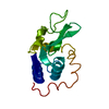
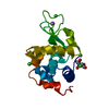
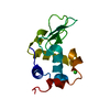
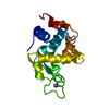
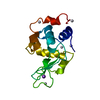
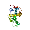
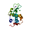
| Title | Chicken egg white lysozyme by highly ordered APA (Anodic Porous Alumina) nanotemplate crystallization method | ||||||
 Components Components | Lysozyme C | ||||||
 Keywords Keywords |  HYDROLASE / Hen Egg White Lysozyme / Allergen / Antimicrobial / Bacteriolytic enzyme / Disulfide bond / Glycosidase HYDROLASE / Hen Egg White Lysozyme / Allergen / Antimicrobial / Bacteriolytic enzyme / Disulfide bond / Glycosidase | ||||||
| Function / homology |  Function and homology informationAntimicrobial peptides / Neutrophil degranulation / beta-N-acetylglucosaminidase activity / cell wall macromolecule catabolic process / lysozyme / lysozyme activity / killing of cells of another organism / defense response to Gram-negative bacterium / defense response to Gram-positive bacterium / defense response to bacterium ...Antimicrobial peptides / Neutrophil degranulation / beta-N-acetylglucosaminidase activity / cell wall macromolecule catabolic process / lysozyme / lysozyme activity / killing of cells of another organism / defense response to Gram-negative bacterium / defense response to Gram-positive bacterium / defense response to bacterium / endoplasmic reticulum / extracellular space / identical protein binding / cytoplasm Function and homology informationAntimicrobial peptides / Neutrophil degranulation / beta-N-acetylglucosaminidase activity / cell wall macromolecule catabolic process / lysozyme / lysozyme activity / killing of cells of another organism / defense response to Gram-negative bacterium / defense response to Gram-positive bacterium / defense response to bacterium ...Antimicrobial peptides / Neutrophil degranulation / beta-N-acetylglucosaminidase activity / cell wall macromolecule catabolic process / lysozyme / lysozyme activity / killing of cells of another organism / defense response to Gram-negative bacterium / defense response to Gram-positive bacterium / defense response to bacterium / endoplasmic reticulum / extracellular space / identical protein binding / cytoplasmSimilarity search - Function | ||||||
| Biological species |  Gallus gallus (chicken) Gallus gallus (chicken) | ||||||
| Method | X-RAY DIFFRACTION / SYNCHROTRON / MOLECULAR REPLACEMENT / molecular replacement / Resolution: 1.6 Å | ||||||
 Authors Authors | Pechkova, E. / Tripathi, S.K. / Nicolini, C. | ||||||
 Citation Citation | Journal: To be Published Title: Comparison of lysozyme crystals grown by APA and classical hanging drop method Authors: Pechkova, E. / Tripathi, S.K. / Nicolini, C. | ||||||
| History |
|
- Structure visualization
| Structure viewer | Molecule: MolmilJmol/JSmol |
|---|
- Downloads & links
-Download
| PDBx/mmCIF format | 3iju.cif.gz | 39.3 KB | Display | PDBx/mmCIF format |
|---|---|---|---|---|
| PDB format | pdb3iju.ent.gz | 26.5 KB | Display | PDB format |
| PDBx/mmJSON format | 3iju.json.gz | Tree view | PDBx/mmJSON format | |
| Others |  Other downloads Other downloads |
-Validation report
| Arichive directory | https://data.pdbj.org/pub/pdb/validation_reports/ij/3ijuftp://data.pdbj.org/pub/pdb/validation_reports/ij/3iju | HTTPS FTP |
|---|
-Related structure data
| Related structure data | 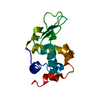 3ijvC  2yvbS S: Starting model for refinement C: citing same article ( |
|---|---|
| Similar structure data |


-Links
 PDBj
PDBj


- Assembly
| Deposited unit | 
| ||||||||
|---|---|---|---|---|---|---|---|---|---|
| 1 |
| ||||||||
| Unit cell |
|
-Components
| #1: Protein | Mass: 14331.160 Da / Num. of mol.: 1 / Source method: isolated from a natural source / Details: Egg / Source: (natural) Gallus gallus (chicken) / Strain: white leghorn / References: UniProt: P00698, lysozyme |
|---|---|
| #2: Water | ChemComp-HOH / Water Mass: 18.015 Da / Num. of mol.: 81 / Source method: isolated from a natural source / Formula: H2O Mass: 18.015 Da / Num. of mol.: 81 / Source method: isolated from a natural source / Formula: H2O |
-Experimental details
-Experiment
| Experiment | Method: X-RAY DIFFRACTION / Number of used crystals: 1 |
|---|
- Sample preparation
| Crystal | Density Matthews: 2.05 Å3/Da / Density % sol: 39.91 % / Mosaicity: 0.42 ° |
|---|---|
| Crystal grow | Temperature: 293 K / Method: vapor diffusion, hanging drop / pH: 4.7 Details: 0.04N NaOAc/HOAc buffer, pH 4.7, VAPOR DIFFUSION, HANGING DROP, temperature 293K |
-Data collection
| Diffraction | Mean temperature: 100 K |
|---|---|
| Diffraction source | Source: SYNCHROTRON / Site: ESRF  / Beamline: ID13 / Wavelength: 0.9465 Å / Beamline: ID13 / Wavelength: 0.9465 Å |
| Detector | Type: MAR CCD 165 mm / Detector: CCD / Date: Aug 31, 2006 / Details: mirror |
| Radiation | Monochromator: Si-111 / Protocol: SINGLE WAVELENGTH / Monochromatic (M) / Laue (L): M / Scattering type: x-ray |
| Radiation wavelength | Wavelength: 0.9465 Å / Relative weight: 1 |
| Reflection | Resolution: 1.6→55.907 Å / Num. obs: 16036 / % possible obs: 98.9 % / Redundancy: 11 % / Rmerge(I) obs: 0.099 / Rsym value: 0.099 / Net I/σ(I): 21.6 / Num. measured all: 176731 |
| Reflection shell | Resolution: 1.6→1.69 Å / Redundancy: 11.5 % / Rmerge(I) obs: 0.221 / Mean I/σ(I) obs: 2.8 / Num. measured all: 26673 / Num. unique all: 2322 / Rsym value: 0.221 / % possible all: 100 |
-Phasing
| Phasing | Method: molecular replacement |
|---|
- Processing
| Software |
| ||||||||||||||||||||||||||||||||||||||||||||||||||||||||||||||||||||||||||||||||||||||||||
|---|---|---|---|---|---|---|---|---|---|---|---|---|---|---|---|---|---|---|---|---|---|---|---|---|---|---|---|---|---|---|---|---|---|---|---|---|---|---|---|---|---|---|---|---|---|---|---|---|---|---|---|---|---|---|---|---|---|---|---|---|---|---|---|---|---|---|---|---|---|---|---|---|---|---|---|---|---|---|---|---|---|---|---|---|---|---|---|---|---|---|---|
| Refinement | Method to determine structure: MOLECULAR REPLACEMENT Starting model: PDB ENTRY 2yvb Resolution: 1.6→21.57 Å / Cor.coef. Fo:Fc: 0.927 / Cor.coef. Fo:Fc free: 0.929 / WRfactor Rfree: 0.265 / WRfactor Rwork: 0.244 / Occupancy max: 1 / Occupancy min: 1 / FOM work R set: 0.845 / SU B: 1.762 / SU ML: 0.065 / SU R Cruickshank DPI: 0.116 / SU Rfree: 0.104 / Cross valid method: THROUGHOUT / σ(F): 0 / ESU R: 0.116 / ESU R Free: 0.104 / Stereochemistry target values: MAXIMUM LIKELIHOOD / Details: HYDROGENS HAVE BEEN ADDED IN THE RIDING POSITIONS
| ||||||||||||||||||||||||||||||||||||||||||||||||||||||||||||||||||||||||||||||||||||||||||
| Solvent computation | Ion probe radii: 0.8 Å / Shrinkage radii: 0.8 Å / VDW probe radii: 1.2 Å / Solvent model: MASK | ||||||||||||||||||||||||||||||||||||||||||||||||||||||||||||||||||||||||||||||||||||||||||
| Displacement parameters | Biso max: 64.53 Å2 / Biso mean: 18.645 Å2 / Biso min: 8.07 Å2
| ||||||||||||||||||||||||||||||||||||||||||||||||||||||||||||||||||||||||||||||||||||||||||
| Refinement step | Cycle: LAST / Resolution: 1.6→21.57 Å
| ||||||||||||||||||||||||||||||||||||||||||||||||||||||||||||||||||||||||||||||||||||||||||
| Refine LS restraints |
| ||||||||||||||||||||||||||||||||||||||||||||||||||||||||||||||||||||||||||||||||||||||||||
| LS refinement shell | Resolution: 1.6→1.641 Å / Total num. of bins used: 20
|
