 Movie
Movie Controller

[English] 日本語
 Yorodumi
- PDB-3ho0: Crystal structure of the PPARgamma-LBD complexed with a new arylo... -
+ Open data

- Basic information
| Entry | Database: PDB / ID: 3ho0 | ||||||
|---|---|---|---|---|---|---|---|
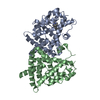
| Title | Crystal structure of the PPARgamma-LBD complexed with a new aryloxy-3phenylpropanoic acid | ||||||
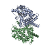
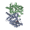
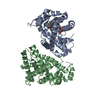
 Components Components | Peroxisome proliferator-activated receptor gamma | ||||||
 Keywords Keywords | TRANSCRIPTION / BUNDLE OF ALPHA-HELICES / SMALL FOUR-STRANDED BETA-SHEET / Activator / Alternative splicing / Diabetes mellitus / Disease mutation / DNA-binding / Metal-binding / Nucleus / Obesity / Phosphoprotein / Polymorphism / Receptor / Transcription regulation / Zinc / Zinc-finger | ||||||
| Function / homology |  Function and homology information Function and homology informationComplex I biogenesis / Respiratory electron transport / mitochondrial ATP synthesis coupled electron transport / mitochondrial respiratory chain complex I assembly / : / mitochondrial electron transport, NADH to ubiquinone / proton motive force-driven mitochondrial ATP synthesis / Mitochondrial protein degradation / NADH dehydrogenase (ubiquinone) activity / mitochondrial membrane ...Complex I biogenesis / Respiratory electron transport / mitochondrial ATP synthesis coupled electron transport / mitochondrial respiratory chain complex I assembly / : / mitochondrial electron transport, NADH to ubiquinone / proton motive force-driven mitochondrial ATP synthesis / Mitochondrial protein degradation / NADH dehydrogenase (ubiquinone) activity / mitochondrial membrane / aerobic respiration / mitochondrial inner membrane / mitochondrion / nucleoplasm Similarity search - Function | ||||||
| Biological species |  Homo sapiens (human) Homo sapiens (human) | ||||||
| Method |  X-RAY DIFFRACTION / SYNCHROTRON / MOLECULAR REPLACEMENT / Resolution: 2.6 Å X-RAY DIFFRACTION / SYNCHROTRON / MOLECULAR REPLACEMENT / Resolution: 2.6 Å | ||||||
 Authors Authors | Pochetti, G. / Montanari, R. / Mazza, F. / Loiodice, F. / Fracchiolla, G. / Laghezza, A. / Lavecchia, A. / Novellino, E. | ||||||
 Citation Citation | Journal: J.Med.Chem. / Year: 2009 Title: New 2-Aryloxy-3-phenyl-propanoic Acids As Peroxisome Proliferator-Activated Receptors alpha/gamma Dual Agonists with Improved Potency and Reduced Adverse Effects on Skeletal Muscle Function Authors: Fracchiolla, G. / Laghezza, A. / Piemontese, L. / Tortorella, P. / Mazza, F. / Montanari, R. / Pochetti, G. / Lavecchia, A. / Novellino, E. / Pierno, S. / Conte Camerino, D. / Loiodice, F. #1: Journal: J.Med.Chem. / Year: 2008Title: Crystal structure of the peroxisome proliferator-activated receptor gamma (PPARgamma) ligand binding domain complexed with a novel partial agonist: a new region of the hydrophobic pocket could ...Title: Crystal structure of the peroxisome proliferator-activated receptor gamma (PPARgamma) ligand binding domain complexed with a novel partial agonist: a new region of the hydrophobic pocket could be exploited for drug design Authors: Montanari, R. / Saccoccia, F. / Scotti, E. / Crestani, M. / Godio, C. / Gilardi, F. / Loiodice, F. / Fracchiolla, G. / Laghezza, A. / Tortorella, P. / Lavecchia, A. / Novellino, E. / Mazza, ...Authors: Montanari, R. / Saccoccia, F. / Scotti, E. / Crestani, M. / Godio, C. / Gilardi, F. / Loiodice, F. / Fracchiolla, G. / Laghezza, A. / Tortorella, P. / Lavecchia, A. / Novellino, E. / Mazza, F. / Aschi, M. / Pochetti, G. | ||||||
| History |
|
- Structure visualization
| Structure viewer | Molecule: MolmilJmol/JSmol |
|---|
- Downloads & links
-Download
| PDBx/mmCIF format | 3ho0.cif.gz | 121 KB | Display | PDBx/mmCIF format |
|---|---|---|---|---|
| PDB format | pdb3ho0.ent.gz | 94.3 KB | Display | PDB format |
| PDBx/mmJSON format | 3ho0.json.gz | Tree view | PDBx/mmJSON format | |
| Others |  Other downloads Other downloads |
-Validation report
| Summary document | 3ho0_validation.pdf.gz | 696.3 KB | Display | wwPDB validaton report |
|---|---|---|---|---|
| Full document | 3ho0_full_validation.pdf.gz | 717.3 KB | Display | |
| Data in XML | 3ho0_validation.xml.gz | 23.7 KB | Display | |
| Data in CIF | 3ho0_validation.cif.gz | 32.4 KB | Display | |
| Arichive directory | https://data.pdbj.org/pub/pdb/validation_reports/ho/3ho0ftp://data.pdbj.org/pub/pdb/validation_reports/ho/3ho0 | HTTPS FTP |
-Related structure data
| Related structure data | 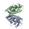 3hodC  3b3kS S: Starting model for refinement C: citing same article ( |
|---|---|
| Similar structure data |


-Links
 PDBj
PDBj- Assembly
| Deposited unit | 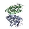
| ||||||||
|---|---|---|---|---|---|---|---|---|---|
| 1 | 
| ||||||||
| Unit cell |
|
-Components
| #1: Protein | Mass: 32530.652 Da / Num. of mol.: 2 / Fragment: ligand binding domain (LBD), UNP residues 223-504 Source method: isolated from a genetically manipulated source Source: (gene. exp.) Homo sapiens (human) / Gene: PPARG, NR1C3 / Plasmid: pET28a / Production host:  #2: Chemical | ChemComp-DKD / ( | 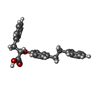  Mass: 346.419 Da / Num. of mol.: 1 / Source method: obtained synthetically / Formula: C23H22O3 Mass: 346.419 Da / Num. of mol.: 1 / Source method: obtained synthetically / Formula: C23H22O3#3: Water | ChemComp-HOH / |  Mass: 18.015 Da / Num. of mol.: 106 / Source method: isolated from a natural source / Formula: H2O Mass: 18.015 Da / Num. of mol.: 106 / Source method: isolated from a natural source / Formula: H2O |
|---|
-Experimental details
-Experiment
| Experiment | Method: X-RAY DIFFRACTION / Number of used crystals: 1 |
|---|
- Sample preparation
| Crystal | Density Matthews: 2.62 Å3/Da / Density % sol: 53.03 % |
|---|---|
| Crystal grow | Temperature: 293 K / Method: vapor diffusion, sitting drop / pH: 8 Details: 0.8M NaCitrate, 0.15M Tris, pH8.0, VAPOR DIFFUSION, SITTING DROP, temperature 293K |
-Data collection
| Diffraction | Mean temperature: 100 K |
|---|---|
| Diffraction source | Source: SYNCHROTRON / Site: ESRF  / Beamline: ID14-2 / Wavelength: 0.933 Å / Beamline: ID14-2 / Wavelength: 0.933 Å |
| Detector | Type: ADSC QUANTUM 4 / Detector: CCD / Date: May 9, 2008 |
| Radiation | Monochromator: Diamond (111), Ge(220) / Protocol: SINGLE WAVELENGTH / Monochromatic (M) / Laue (L): M / Scattering type: x-ray |
| Radiation wavelength | Wavelength: 0.933 Å / Relative weight: 1 |
| Reflection | Resolution: 2.6→25 Å / Num. obs: 20618 / % possible obs: 5 % / Observed criterion σ(F): 0 / Observed criterion σ(I): 0 / Redundancy: 3.8 % / Biso Wilson estimate: 68.4 Å2 / Rmerge(I) obs: 0.083 / Net I/σ(I): 4.8 |
| Reflection shell | Resolution: 2.6→2.74 Å / Redundancy: 3.9 % / Rmerge(I) obs: 0.349 / Mean I/σ(I) obs: 1.9 / % possible all: 99 |
- Processing
| Software |
| ||||||||||||||||||||
|---|---|---|---|---|---|---|---|---|---|---|---|---|---|---|---|---|---|---|---|---|---|
| Refinement | Method to determine structure: MOLECULAR REPLACEMENT Starting model: PDB ENTRY 3B3K Resolution: 2.6→10 Å / Isotropic thermal model: Isotropic / σ(F): 0 / Stereochemistry target values: Engh & Huber
| ||||||||||||||||||||
| Refinement step | Cycle: LAST / Resolution: 2.6→10 Å
| ||||||||||||||||||||
| Refine LS restraints |
|
