 Movie
Movie Controller


+ Open data

- Basic information
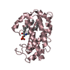
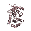
| Entry | Database: PDB / ID: 3hcm | ||||||
|---|---|---|---|---|---|---|---|
| Title | Crystal structure of human S100B in complex with S45 | ||||||
 Components Components | Protein S100-B | ||||||
 Keywords Keywords | METAL BINDING PROTEIN /  S100B / calcium binding protein / inhibitor / Calcium / Cytoplasm / Metal-binding / Nucleus S100B / calcium binding protein / inhibitor / Calcium / Cytoplasm / Metal-binding / Nucleus | ||||||
| Function / homology |  Function and homology informationadaptive thermogenesis / sympathetic neuron projection extension / RAGE receptor binding / ion binding / S100 protein binding / regulation of neuronal synaptic plasticity / TRAF6 mediated NF-kB activation / Advanced glycosylation endproduct receptor signaling / Nuclear signaling by ERBB4 / ruffle ...adaptive thermogenesis / sympathetic neuron projection extension / RAGE receptor binding / ion binding / S100 protein binding / regulation of neuronal synaptic plasticity / TRAF6 mediated NF-kB activation / Advanced glycosylation endproduct receptor signaling / Nuclear signaling by ERBB4 / ruffle / positive regulation of neuron differentiation / axonogenesis / central nervous system development / tau protein binding / TAK1-dependent IKK and NF-kappa-B activation / memory / calcium-dependent protein binding / positive regulation of canonical NF-kappaB signal transduction / learning or memory / cell adhesion / intracellular membrane-bounded organelle / neuronal cell body / calcium ion binding / positive regulation of cell population proliferation / perinuclear region of cytoplasm / protein homodimerization activity / extracellular space / zinc ion binding / extracellular region / nucleoplasm / identical protein binding / nucleus / cytosol / cytoplasm Function and homology informationadaptive thermogenesis / sympathetic neuron projection extension / RAGE receptor binding / ion binding / S100 protein binding / regulation of neuronal synaptic plasticity / TRAF6 mediated NF-kB activation / Advanced glycosylation endproduct receptor signaling / Nuclear signaling by ERBB4 / ruffle ...adaptive thermogenesis / sympathetic neuron projection extension / RAGE receptor binding / ion binding / S100 protein binding / regulation of neuronal synaptic plasticity / TRAF6 mediated NF-kB activation / Advanced glycosylation endproduct receptor signaling / Nuclear signaling by ERBB4 / ruffle / positive regulation of neuron differentiation / axonogenesis / central nervous system development / tau protein binding / TAK1-dependent IKK and NF-kappa-B activation / memory / calcium-dependent protein binding / positive regulation of canonical NF-kappaB signal transduction / learning or memory / cell adhesion / intracellular membrane-bounded organelle / neuronal cell body / calcium ion binding / positive regulation of cell population proliferation / perinuclear region of cytoplasm / protein homodimerization activity / extracellular space / zinc ion binding / extracellular region / nucleoplasm / identical protein binding / nucleus / cytosol / cytoplasmSimilarity search - Function | ||||||
| Biological species |  Homo sapiens (human) Homo sapiens (human) | ||||||
| Method | X-RAY DIFFRACTION / SYNCHROTRON / MOLECULAR REPLACEMENT / Resolution: 2 Å | ||||||
 Authors Authors | Mangani, S. / Cesari, L. | ||||||
 Citation Citation | Journal: Chemmedchem / Year: 2010 Title: Fragmenting the S100B-p53 Interaction: Combined Virtual/Biophysical Screening Approaches to Identify Ligands Authors: Agamennone, M. / Cesari, L. / Lalli, D. / Turlizzi, E. / Del Conte, R. / Turano, P. / Mangani, S. / Padova, A. | ||||||
| History |
|
- Structure visualization
| Structure viewer | Molecule: MolmilJmol/JSmol |
|---|
- Downloads & links
-Download
| PDBx/mmCIF format | 3hcm.cif.gz | 54.5 KB | Display | PDBx/mmCIF format |
|---|---|---|---|---|
| PDB format | pdb3hcm.ent.gz | 38.6 KB | Display | PDB format |
| PDBx/mmJSON format | 3hcm.json.gz | Tree view | PDBx/mmJSON format | |
| Others |  Other downloads Other downloads |
-Validation report
| Arichive directory | https://data.pdbj.org/pub/pdb/validation_reports/hc/3hcmftp://data.pdbj.org/pub/pdb/validation_reports/hc/3hcm | HTTPS FTP |
|---|
-Related structure data
| Related structure data |  2h61S S: Starting model for refinement |
|---|---|
| Similar structure data |


-Links
 PDBj
PDBj


- Assembly
| Deposited unit | 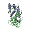
| ||||||||
|---|---|---|---|---|---|---|---|---|---|
| 1 |
| ||||||||
| Unit cell |
|
-Components
| #1: Protein | Mass: 10727.037 Da / Num. of mol.: 2 Source method: isolated from a genetically manipulated source Source: (gene. exp.) Homo sapiens (human)Description: The protein was purchased from a commercial source. Production host:  Escherichia coli (E. coli) / References: UniProt: P04271 Escherichia coli (E. coli) / References: UniProt: P04271#2: Chemical | ChemComp-CA /   Mass: 40.078 Da / Num. of mol.: 4 / Source method: obtained synthetically / Formula: Ca Mass: 40.078 Da / Num. of mol.: 4 / Source method: obtained synthetically / Formula: Ca#3: Chemical | 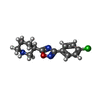  Mass: 263.723 Da / Num. of mol.: 2 / Source method: obtained synthetically / Formula: C13H14ClN3O Mass: 263.723 Da / Num. of mol.: 2 / Source method: obtained synthetically / Formula: C13H14ClN3O#4: Chemical | ChemComp-ACT / | Acetate  Mass: 59.044 Da / Num. of mol.: 1 / Source method: obtained synthetically / Formula: C2H3O2 Mass: 59.044 Da / Num. of mol.: 1 / Source method: obtained synthetically / Formula: C2H3O2#5: Water | ChemComp-HOH / | Water Mass: 18.015 Da / Num. of mol.: 96 / Source method: isolated from a natural source / Formula: H2O Mass: 18.015 Da / Num. of mol.: 96 / Source method: isolated from a natural source / Formula: H2O |
|---|
-Experimental details
-Experiment
| Experiment | Method: X-RAY DIFFRACTION / Number of used crystals: 1 |
|---|
- Sample preparation
| Crystal | Density Matthews: 2.21 Å3/Da / Density % sol: 44.31 % |
|---|---|
| Crystal grow | Temperature: 298 K / Method: vapor diffusion, sitting drop / pH: 4.6 Details: 0.1M sodium acetate, 0.02M calcium chloride, 45% methylpenthanediol, pH4.6, VAPOR DIFFUSION, SITTING DROP, temperature 298K |
-Data collection
| Diffraction | Mean temperature: 100 K |
|---|---|
| Diffraction source | Source: SYNCHROTRON / Site: EMBL/DESY, HAMBURG  / Beamline: X11 / Wavelength: 0.814 Å / Beamline: X11 / Wavelength: 0.814 Å |
| Detector | Type: MAR555 FLAT PANEL / Detector: IMAGE PLATE / Date: Oct 24, 2006 |
| Radiation | Monochromator: Si [111] / Protocol: SINGLE WAVELENGTH / Monochromatic (M) / Laue (L): M / Scattering type: x-ray |
| Radiation wavelength | Wavelength: 0.814 Å / Relative weight: 1 |
| Reflection | Resolution: 2→17.68 Å / Num. all: 12148 / Num. obs: 9406 / % possible obs: 81.5 % / Observed criterion σ(F): 0 / Observed criterion σ(I): 2 / Redundancy: 4.3 % / Biso Wilson estimate: 29.93 Å2 / Rsym value: 0.041 / Net I/σ(I): 11.5 |
| Reflection shell | Resolution: 2→2.11 Å / Redundancy: 4.1 % / Rmerge(I) obs: 0.075 / Mean I/σ(I) obs: 9.1 / Num. unique all: 1337 / % possible all: 81.5 |
- Processing
| Software |
| ||||||||||||||||||||||||||||||||||||||||||||||||||||||||||||||||||||||||||||||||||||||||||||||||||||||||||||||||||||||||||||||||||||||||||||||||||||||||||||||||||||||||||
|---|---|---|---|---|---|---|---|---|---|---|---|---|---|---|---|---|---|---|---|---|---|---|---|---|---|---|---|---|---|---|---|---|---|---|---|---|---|---|---|---|---|---|---|---|---|---|---|---|---|---|---|---|---|---|---|---|---|---|---|---|---|---|---|---|---|---|---|---|---|---|---|---|---|---|---|---|---|---|---|---|---|---|---|---|---|---|---|---|---|---|---|---|---|---|---|---|---|---|---|---|---|---|---|---|---|---|---|---|---|---|---|---|---|---|---|---|---|---|---|---|---|---|---|---|---|---|---|---|---|---|---|---|---|---|---|---|---|---|---|---|---|---|---|---|---|---|---|---|---|---|---|---|---|---|---|---|---|---|---|---|---|---|---|---|---|---|---|---|---|---|---|
| Refinement | Method to determine structure: MOLECULAR REPLACEMENT Starting model: PDB ENTRY 2H61 Resolution: 2→17.63 Å / Cor.coef. Fo:Fc: 0.913 / Cor.coef. Fo:Fc free: 0.84 / SU B: 5.87 / SU ML: 0.173 / Cross valid method: THROUGHOUT / σ(F): 0 / σ(I): 2 / ESU R: 0.365 / ESU R Free: 0.281 / Stereochemistry target values: MAXIMUM LIKELIHOOD / Details: HYDROGENS HAVE BEEN ADDED IN THE RIDING POSITIONS
| ||||||||||||||||||||||||||||||||||||||||||||||||||||||||||||||||||||||||||||||||||||||||||||||||||||||||||||||||||||||||||||||||||||||||||||||||||||||||||||||||||||||||||
| Solvent computation | Ion probe radii: 0.8 Å / Shrinkage radii: 0.8 Å / VDW probe radii: 1.2 Å / Solvent model: MASK | ||||||||||||||||||||||||||||||||||||||||||||||||||||||||||||||||||||||||||||||||||||||||||||||||||||||||||||||||||||||||||||||||||||||||||||||||||||||||||||||||||||||||||
| Displacement parameters | Biso mean: 33.961 Å2
| ||||||||||||||||||||||||||||||||||||||||||||||||||||||||||||||||||||||||||||||||||||||||||||||||||||||||||||||||||||||||||||||||||||||||||||||||||||||||||||||||||||||||||
| Refinement step | Cycle: LAST / Resolution: 2→17.63 Å
| ||||||||||||||||||||||||||||||||||||||||||||||||||||||||||||||||||||||||||||||||||||||||||||||||||||||||||||||||||||||||||||||||||||||||||||||||||||||||||||||||||||||||||
| Refine LS restraints |
| ||||||||||||||||||||||||||||||||||||||||||||||||||||||||||||||||||||||||||||||||||||||||||||||||||||||||||||||||||||||||||||||||||||||||||||||||||||||||||||||||||||||||||
| LS refinement shell | Resolution: 2→2.051 Å / Total num. of bins used: 20
|
