| 登録情報 | データベース: PDB / ID: 3fw6
|
|---|
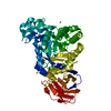
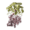
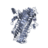
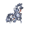
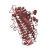
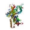
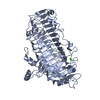
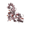
| タイトル | Crystal structure of CelM2, a bifunctional glucanase-xylanase protein from a metagenome library |
|---|
 要素 要素 | Cellulase |
|---|
 キーワード キーワード | HYDROLASE / CelM2 / glucanase-xyanase / glucanase / xylanase / bifunctional enzyme |
|---|
| 機能・相同性 |  機能・相同性情報 機能・相同性情報
Glycoside hydrolase, family 44 / Glycoside hydrolase family 44 / PKD domain / Polycystic kidney disease (PKD) domain profile. / PKD domain / PKD domain superfamily / PKD/Chitinase domain / Repeats in polycystic kidney disease 1 (PKD1) and other proteins / Golgi alpha-mannosidase II / Glycosyl hydrolase, all-beta ...Glycoside hydrolase, family 44 / Glycoside hydrolase family 44 / PKD domain / Polycystic kidney disease (PKD) domain profile. / PKD domain / PKD domain superfamily / PKD/Chitinase domain / Repeats in polycystic kidney disease 1 (PKD1) and other proteins / Golgi alpha-mannosidase II / Glycosyl hydrolase, all-beta / Glycosidases / Glycoside hydrolase superfamily / TIM Barrel / Alpha-Beta Barrel / Immunoglobulin-like fold / Immunoglobulin-like / Sandwich / Mainly Beta / Alpha Beta類似検索 - ドメイン・相同性 |
|---|
| 生物種 |  uncultured bacterium (環境試料) uncultured bacterium (環境試料) |
|---|
| 手法 |  X線回折 / シンクロトロン / 分子置換 / 解像度: 2.3 Å X線回折 / シンクロトロン / 分子置換 / 解像度: 2.3 Å |
|---|
 データ登録者 データ登録者 | Hwang, K.Y. / Nam, K.H. |
|---|
 引用 引用 | ジャーナル: Biochem.Biophys.Res.Commun. / 年: 2009
タイトル: Crystal structure of CelM2, a bifunctional glucanase-xylanase protein from a metagenome library
著者: Nam, K.H. / Kim, S.-J. / Hwang, K.Y. |
|---|
| 履歴 | | 登録 | 2009年1月17日 | 登録サイト: RCSB / 処理サイト: PDBJ |
|---|
| 改定 1.0 | 2009年3月3日 | Provider: repository / タイプ: Initial release |
|---|
| 改定 1.1 | 2011年7月13日 | Group: Version format compliance |
|---|
| 改定 1.2 | 2023年11月1日 | Group: Data collection / Database references ...Data collection / Database references / Derived calculations / Refinement description
カテゴリ: chem_comp_atom / chem_comp_bond ...chem_comp_atom / chem_comp_bond / database_2 / pdbx_initial_refinement_model / struct_conn / struct_ref_seq_dif / struct_site
Item: _database_2.pdbx_DOI / _database_2.pdbx_database_accession ..._database_2.pdbx_DOI / _database_2.pdbx_database_accession / _struct_conn.ptnr1_auth_comp_id / _struct_conn.ptnr1_auth_seq_id / _struct_conn.ptnr1_label_asym_id / _struct_conn.ptnr1_label_atom_id / _struct_conn.ptnr1_label_comp_id / _struct_conn.ptnr1_label_seq_id / _struct_conn.ptnr2_auth_comp_id / _struct_conn.ptnr2_auth_seq_id / _struct_conn.ptnr2_label_asym_id / _struct_conn.ptnr2_label_atom_id / _struct_conn.ptnr2_label_comp_id / _struct_conn.ptnr2_label_seq_id / _struct_ref_seq_dif.details / _struct_site.pdbx_auth_asym_id / _struct_site.pdbx_auth_comp_id / _struct_site.pdbx_auth_seq_id |
|---|
|
|---|
 ムービー
ムービー コントローラー


 データを開く
 基本情報
基本情報 構造の表示
構造の表示 ダウンロードとリンク
 その他のダウンロード
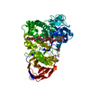


 PDBj
PDBj 集合体
集合体

 分子量: 65.409 Da / 分子数: 3 / 由来タイプ: 合成 / 式: Zn
 分子量: 18.015 Da / 分子数: 252 / 由来タイプ: 天然 / 式: H2O
 試料調製
試料調製 / ビームライン: 4A / 波長: 1 Å
 解析
解析