 Movie
Movie Controller


+ Open data

- Basic information
| Entry | Database: PDB / ID: 3fck | ||||||
|---|---|---|---|---|---|---|---|
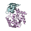
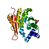
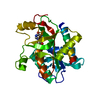
| Title | Complex of UNG2 and a fragment-based design inhibitor | ||||||
 Components Components | Uracil-DNA glycosylase | ||||||
 Keywords Keywords | HYDROLASE / DNA REPAIR / URACIL / URACIL DNA GLYCOSYLASE / Alternative splicing / Disease mutation / DNA damage / Glycosidase / Host-virus interaction / Mitochondrion / Nucleus / Phosphoprotein / Transit peptide | ||||||
| Function / homology |  Function and homology informationbase-excision repair, AP site formation via deaminated base removal / uracil-DNA glycosylase / depyrimidination / Displacement of DNA glycosylase by APEX1 / isotype switching / uracil DNA N-glycosylase activity / ribosomal small subunit binding / somatic hypermutation of immunoglobulin genes / Recognition and association of DNA glycosylase with site containing an affected pyrimidine / Cleavage of the damaged pyrimidine ...base-excision repair, AP site formation via deaminated base removal / uracil-DNA glycosylase / depyrimidination / Displacement of DNA glycosylase by APEX1 / isotype switching / uracil DNA N-glycosylase activity / ribosomal small subunit binding / somatic hypermutation of immunoglobulin genes / Recognition and association of DNA glycosylase with site containing an affected pyrimidine / Cleavage of the damaged pyrimidine / Chromatin modifications during the maternal to zygotic transition (MZT) / base-excision repair / damaged DNA binding / negative regulation of apoptotic process / mitochondrion / nucleoplasm / nucleus Function and homology informationbase-excision repair, AP site formation via deaminated base removal / uracil-DNA glycosylase / depyrimidination / Displacement of DNA glycosylase by APEX1 / isotype switching / uracil DNA N-glycosylase activity / ribosomal small subunit binding / somatic hypermutation of immunoglobulin genes / Recognition and association of DNA glycosylase with site containing an affected pyrimidine / Cleavage of the damaged pyrimidine ...base-excision repair, AP site formation via deaminated base removal / uracil-DNA glycosylase / depyrimidination / Displacement of DNA glycosylase by APEX1 / isotype switching / uracil DNA N-glycosylase activity / ribosomal small subunit binding / somatic hypermutation of immunoglobulin genes / Recognition and association of DNA glycosylase with site containing an affected pyrimidine / Cleavage of the damaged pyrimidine / Chromatin modifications during the maternal to zygotic transition (MZT) / base-excision repair / damaged DNA binding / negative regulation of apoptotic process / mitochondrion / nucleoplasm / nucleusSimilarity search - Function | ||||||
| Biological species |  Homo sapiens (human) Homo sapiens (human) | ||||||
| Method | X-RAY DIFFRACTION / MOLECULAR REPLACEMENT / molecular replacement / Resolution: 1.64 Å | ||||||
 Authors Authors | Bianchet, M.A. / Chung, S. / Parker, J.B. / Amzel, L.M. / Stivers, J.T. | ||||||
 Citation Citation | Journal: Nat.Chem.Biol. / Year: 2009 Title: Impact of linker strain and flexibility in the design of a fragment-based inhibitor Authors: Chung, S. / Parker, J.B. / Bianchet, M. / Amzel, L.M. / Stivers, J.T. #1: Journal: J.Am.Chem.Soc. / Year: 2005Title: Uracil-directed ligand tethering: an efficient strategy for uracil DNA glycosylase (UNG) inhibitor development Authors: Jiang, T.L. / Krosky, D.J. / Seiple, L. / Stivers, J.T. #2: Journal: Nucleic Acids Res. / Year: 2006Title: Mimicking damaged DNA with a small molecule inhibitor of human UNG2. Authors: Krosky, D.J. / Bianchet, M.A. / Seiple, L. / Chung, S. / Amzel, L.M. / Stivers, J.T. | ||||||
| History |
|
- Structure visualization
| Structure viewer | Molecule: MolmilJmol/JSmol |
|---|
- Downloads & links
-Download
| PDBx/mmCIF format | 3fck.cif.gz | 64.4 KB | Display | PDBx/mmCIF format |
|---|---|---|---|---|
| PDB format | pdb3fck.ent.gz | 46 KB | Display | PDB format |
| PDBx/mmJSON format | 3fck.json.gz | Tree view | PDBx/mmJSON format | |
| Others |  Other downloads Other downloads |
-Validation report
| Arichive directory | https://data.pdbj.org/pub/pdb/validation_reports/fc/3fckftp://data.pdbj.org/pub/pdb/validation_reports/fc/3fck | HTTPS FTP |
|---|
-Related structure data


-Links
 PDBj
PDBj
- Assembly
| Deposited unit | 
| ||||||||
|---|---|---|---|---|---|---|---|---|---|
| 1 |
| ||||||||
| Unit cell |
|
-Components
| #1: Protein | / UDG Mass: 25544.137 Da / Num. of mol.: 1 Source method: isolated from a genetically manipulated source Source: (gene. exp.) Homo sapiens (human) / Gene: UNG, DGU, UNG1, UNG15 / Production host:  ESCHERICHIA COLI (E. coli) ESCHERICHIA COLI (E. coli)References: UniProt: P13051, Hydrolases; Glycosylases; Hydrolysing N-glycosyl compounds |
|---|---|
| #2: Chemical | ChemComp-FCK /   Mass: 346.338 Da / Num. of mol.: 1 / Source method: obtained synthetically / Formula: C16H18N4O5 Mass: 346.338 Da / Num. of mol.: 1 / Source method: obtained synthetically / Formula: C16H18N4O5 |
| #3: Water | ChemComp-HOH / Water Mass: 18.015 Da / Num. of mol.: 210 / Source method: isolated from a natural source / Formula: H2O Mass: 18.015 Da / Num. of mol.: 210 / Source method: isolated from a natural source / Formula: H2O |
-Experimental details
-Experiment
| Experiment | Method: X-RAY DIFFRACTION / Number of used crystals: 1 |
|---|
- Sample preparation
| Crystal | Density Matthews: 2.05 Å3/Da / Density % sol: 39.9 % |
|---|---|
| Crystal grow | Temperature: 295 K / Method: vapor diffusion, hanging drop / pH: 8 Details: 1 MICROLITER OF THE SOLUTION: 0.001 M UNG2 0.05 M TRIS-OAC pH 7.0, 0.15 M NACL 0.001 M DTT 0.005 M INHIBITOR, WAS MIXED WITH EQUAL AMOUNT OF THE SOLUTION CONTAINING 0.16 M KSCN AND 22% PEG ...Details: 1 MICROLITER OF THE SOLUTION: 0.001 M UNG2 0.05 M TRIS-OAC pH 7.0, 0.15 M NACL 0.001 M DTT 0.005 M INHIBITOR, WAS MIXED WITH EQUAL AMOUNT OF THE SOLUTION CONTAINING 0.16 M KSCN AND 22% PEG 3350, VAPOR DIFFUSION, HANGING DROP, temperature 295K |
-Data collection
| Diffraction source | Source: ROTATING ANODE / Type: RIGAKU RUH3R / Wavelength: 1.5418 Å | ||||||||||||||||||||||||||||||||||||||||||||||||||||||||||||||||||
|---|---|---|---|---|---|---|---|---|---|---|---|---|---|---|---|---|---|---|---|---|---|---|---|---|---|---|---|---|---|---|---|---|---|---|---|---|---|---|---|---|---|---|---|---|---|---|---|---|---|---|---|---|---|---|---|---|---|---|---|---|---|---|---|---|---|---|---|
| Detector | Type: RIGAKU RAXIS IV / Detector: IMAGE PLATE / Date: Oct 26, 2007 | ||||||||||||||||||||||||||||||||||||||||||||||||||||||||||||||||||
| Radiation | Protocol: SINGLE WAVELENGTH / Monochromatic (M) / Laue (L): M / Scattering type: x-ray | ||||||||||||||||||||||||||||||||||||||||||||||||||||||||||||||||||
| Radiation wavelength | Wavelength: 1.5418 Å / Relative weight: 1 | ||||||||||||||||||||||||||||||||||||||||||||||||||||||||||||||||||
| Reflection | Resolution: 1.63→50 Å / Num. obs: 22828 / % possible obs: 84.9 % / Redundancy: 4.3 % / Rmerge(I) obs: 0.042 / Χ2: 2.268 | ||||||||||||||||||||||||||||||||||||||||||||||||||||||||||||||||||
| Reflection shell |
|
-Phasing
| Phasing | Method: molecular replacement |
|---|
- Processing
| Software |
| ||||||||||||||||||||||||||||||||||||||||||||||||||||||||||||||||||||||||||||||||||||||||||
|---|---|---|---|---|---|---|---|---|---|---|---|---|---|---|---|---|---|---|---|---|---|---|---|---|---|---|---|---|---|---|---|---|---|---|---|---|---|---|---|---|---|---|---|---|---|---|---|---|---|---|---|---|---|---|---|---|---|---|---|---|---|---|---|---|---|---|---|---|---|---|---|---|---|---|---|---|---|---|---|---|---|---|---|---|---|---|---|---|---|---|---|
| Refinement | Method to determine structure: MOLECULAR REPLACEMENT / Resolution: 1.64→27.2 Å / Cor.coef. Fo:Fc: 0.958 / Cor.coef. Fo:Fc free: 0.945 / WRfactor Rfree: 0.249 / WRfactor Rwork: 0.205 / Occupancy max: 1 / Occupancy min: 0 / FOM work R set: 0.844 / SU B: 2.271 / SU ML: 0.078 / SU R Cruickshank DPI: 0.132 / SU Rfree: 0.123 / Cross valid method: THROUGHOUT / σ(F): 0 / ESU R: 0.132 / ESU R Free: 0.123 / Stereochemistry target values: MAXIMUM LIKELIHOOD / Details: HYDROGENS HAVE BEEN ADDED IN THE RIDING POSITIONS
| ||||||||||||||||||||||||||||||||||||||||||||||||||||||||||||||||||||||||||||||||||||||||||
| Solvent computation | Ion probe radii: 0.8 Å / Shrinkage radii: 0.8 Å / VDW probe radii: 1.2 Å / Solvent model: MASK | ||||||||||||||||||||||||||||||||||||||||||||||||||||||||||||||||||||||||||||||||||||||||||
| Displacement parameters | Biso max: 67.18 Å2 / Biso mean: 25.738 Å2 / Biso min: 2 Å2
| ||||||||||||||||||||||||||||||||||||||||||||||||||||||||||||||||||||||||||||||||||||||||||
| Refinement step | Cycle: LAST / Resolution: 1.64→27.2 Å
| ||||||||||||||||||||||||||||||||||||||||||||||||||||||||||||||||||||||||||||||||||||||||||
| Refine LS restraints |
| ||||||||||||||||||||||||||||||||||||||||||||||||||||||||||||||||||||||||||||||||||||||||||
| LS refinement shell | Resolution: 1.64→1.678 Å / Total num. of bins used: 20
|
