 Movie
Movie Controller


+ Open data

- Basic information
| Entry | Database: PDB / ID: 3cfh | ||||||
|---|---|---|---|---|---|---|---|
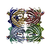
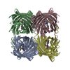
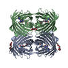
| Title | Photoswitchable red fluorescent protein psRFP, off-state | ||||||
 Components Components | (GFP-like photoswitchable fluorescent protein) x 2 | ||||||
 Keywords Keywords | FLUORESCENT PROTEIN / beta barrel / beta can / chromophore / photoactivation | ||||||
| Function / homology |  Function and homology information Function and homology information | ||||||
| Biological species |  Anemonia sulcata (snake-locks sea anemone) Anemonia sulcata (snake-locks sea anemone) | ||||||
| Method |  X-RAY DIFFRACTION / SYNCHROTRON / MOLECULAR REPLACEMENT / Resolution: 1.75 Å X-RAY DIFFRACTION / SYNCHROTRON / MOLECULAR REPLACEMENT / Resolution: 1.75 Å | ||||||
 Authors Authors | Kachalova, G.S. / Gundel, S. / Bartunik, H.D. / Wiedenmann, J. | ||||||
 Citation Citation | Journal: To be Published Title: Structure of red fluorescent protein asRFP Authors: Gundel, S. / Kachalova, G.S. / Oswald, F. / Fuchs, J. / Bartunik, H.D. / Nienhaus, G.U. / Wiedenmann, J. | ||||||
| History |
|
- Structure visualization
| Structure viewer | Molecule: MolmilJmol/JSmol |
|---|
- Downloads & links
-Download
| PDBx/mmCIF format | 3cfh.cif.gz | 213.7 KB | Display | PDBx/mmCIF format |
|---|---|---|---|---|
| PDB format | pdb3cfh.ent.gz | 174.1 KB | Display | PDB format |
| PDBx/mmJSON format | 3cfh.json.gz | Tree view | PDBx/mmJSON format | |
| Others |  Other downloads Other downloads |
-Validation report
| Summary document | 3cfh_validation.pdf.gz | 496.1 KB | Display | wwPDB validaton report |
|---|---|---|---|---|
| Full document | 3cfh_full_validation.pdf.gz | 527.5 KB | Display | |
| Data in XML | 3cfh_validation.xml.gz | 51.1 KB | Display | |
| Data in CIF | 3cfh_validation.cif.gz | 71.2 KB | Display | |
| Arichive directory | https://data.pdbj.org/pub/pdb/validation_reports/cf/3cfhftp://data.pdbj.org/pub/pdb/validation_reports/cf/3cfh | HTTPS FTP |
-Related structure data
| Related structure data |  3cfaS S: Starting model for refinement |
|---|---|
| Similar structure data |


-Links
 PDBj
PDBj

- Assembly
| Deposited unit | 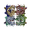
| ||||||||
|---|---|---|---|---|---|---|---|---|---|
| 1 |
| ||||||||
| Unit cell |
| ||||||||
| Details | This protein consists of 4 protein chains (1-231) and inside each of them there is break between 62 and 66 as chromophore. The biological unit is tetramer. |
-Components
| #1: Protein | Mass: 6744.721 Da / Num. of mol.: 4 Source method: isolated from a genetically manipulated source Source: (gene. exp.) Anemonia sulcata (snake-locks sea anemone)Plasmid: pET17b / Production host:  #2: Protein | Mass: 19188.787 Da / Num. of mol.: 4 / Mutation: S143G Source method: isolated from a genetically manipulated source Source: (gene. exp.) Anemonia sulcata (snake-locks sea anemone)Plasmid: pET17b / Production host: #3: Water | ChemComp-HOH / |  Mass: 18.015 Da / Num. of mol.: 908 / Source method: isolated from a natural source / Formula: H2O Mass: 18.015 Da / Num. of mol.: 908 / Source method: isolated from a natural source / Formula: H2O |
|---|
-Experimental details
-Experiment
| Experiment | Method: X-RAY DIFFRACTION / Number of used crystals: 1 |
|---|
- Sample preparation
| Crystal | Density Matthews: 2.79 Å3/Da / Density % sol: 55.9 % |
|---|---|
| Crystal grow | Temperature: 298 K / Method: vapor diffusion, sitting drop Details: 0.1M TRIS, 65%(w/v) MPD, VAPOR DIFFUSION, SITTING DROP, temperature 298.0K |
-Data collection
| Diffraction | Mean temperature: 100 K |
|---|---|
| Diffraction source | Source: SYNCHROTRON / Site: MPG/DESY, HAMBURG  / Beamline: BW6 / Wavelength: 1.05 Å / Beamline: BW6 / Wavelength: 1.05 Å |
| Detector | Type: MAR CCD 165 mm / Detector: CCD / Date: Nov 20, 2007 / Details: mirror |
| Radiation | Monochromator: graphite / Protocol: SINGLE WAVELENGTH / Monochromatic (M) / Laue (L): M / Scattering type: x-ray |
| Radiation wavelength | Wavelength: 1.05 Å / Relative weight: 1 |
| Reflection | Resolution: 1.75→20 Å / Num. all: 115913 / Num. obs: 115191 / % possible obs: 99.4 % / Observed criterion σ(I): 2 / Redundancy: 4.3 % / Biso Wilson estimate: 32.3 Å2 / Rmerge(I) obs: 0.073 / Net I/σ(I): 19.1 |
| Reflection shell | Resolution: 1.75→1.78 Å / Redundancy: 4 % / Rmerge(I) obs: 0.82 / Mean I/σ(I) obs: 2 / Num. unique all: 5751 / % possible all: 99.5 |
- Processing
| Software |
| ||||||||||||||||||||||||||||||||||||||||||||||||||||||||||||||||||||||||||||||||||||||||||
|---|---|---|---|---|---|---|---|---|---|---|---|---|---|---|---|---|---|---|---|---|---|---|---|---|---|---|---|---|---|---|---|---|---|---|---|---|---|---|---|---|---|---|---|---|---|---|---|---|---|---|---|---|---|---|---|---|---|---|---|---|---|---|---|---|---|---|---|---|---|---|---|---|---|---|---|---|---|---|---|---|---|---|---|---|---|---|---|---|---|---|---|
| Refinement | Method to determine structure: MOLECULAR REPLACEMENT Starting model: PDB ENTRY 3CFA Resolution: 1.75→12.27 Å / Cor.coef. Fo:Fc: 0.965 / Cor.coef. Fo:Fc free: 0.953 / SU B: 2.849 / SU ML: 0.09 / Cross valid method: THROUGHOUT / ESU R: 0.121 / ESU R Free: 0.114 / Stereochemistry target values: MAXIMUM LIKELIHOOD
| ||||||||||||||||||||||||||||||||||||||||||||||||||||||||||||||||||||||||||||||||||||||||||
| Solvent computation | Ion probe radii: 0.8 Å / Shrinkage radii: 0.8 Å / VDW probe radii: 1.4 Å / Solvent model: BABINET MODEL WITH MASK | ||||||||||||||||||||||||||||||||||||||||||||||||||||||||||||||||||||||||||||||||||||||||||
| Displacement parameters | Biso mean: 39.785 Å2
| ||||||||||||||||||||||||||||||||||||||||||||||||||||||||||||||||||||||||||||||||||||||||||
| Refinement step | Cycle: LAST / Resolution: 1.75→12.27 Å
| ||||||||||||||||||||||||||||||||||||||||||||||||||||||||||||||||||||||||||||||||||||||||||
| Refine LS restraints |
| ||||||||||||||||||||||||||||||||||||||||||||||||||||||||||||||||||||||||||||||||||||||||||
| LS refinement shell | Resolution: 1.75→1.795 Å / Total num. of bins used: 20
|
