 Movie
Movie Controller


+ Open data

- Basic information
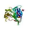
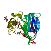
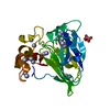
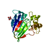
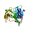
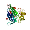
| Entry | Database: PDB / ID: 3ald | ||||||
|---|---|---|---|---|---|---|---|
| Title | Crystal structure of sweet-tasting protein Thaumatin I at 1.10 A | ||||||
 Components Components | Thaumatin I | ||||||
 Keywords Keywords | PLANT PROTEIN / THAUMATIN / SWEET-TASTING PROTEIN | ||||||
| Function / homology |  Function and homology information Function and homology information | ||||||
| Biological species |  Thaumatococcus daniellii (katemfe) Thaumatococcus daniellii (katemfe) | ||||||
| Method | X-RAY DIFFRACTION / SYNCHROTRON / AB INITIO / Resolution: 1.1 Å | ||||||
 Authors Authors | Masuda, T. / Mikami, B. / Kitabatake, N. | ||||||
 Citation Citation | Journal: Acta Crystallogr.,Sect.F / Year: 2011 Title: High-resolution structure of the recombinant sweet-tasting protein thaumatin I Authors: Masuda, T. / Ohta, K. / Mikami, B. / Kitabatake, N. | ||||||
| History |
|
- Structure visualization
| Structure viewer | Molecule: MolmilJmol/JSmol |
|---|
- Downloads & links
-Download
| PDBx/mmCIF format | 3ald.cif.gz | 146.8 KB | Display | PDBx/mmCIF format |
|---|---|---|---|---|
| PDB format | pdb3ald.ent.gz | 126 KB | Display | PDB format |
| PDBx/mmJSON format | 3ald.json.gz | Tree view | PDBx/mmJSON format | |
| Others |  Other downloads Other downloads |
-Validation report
| Arichive directory | https://data.pdbj.org/pub/pdb/validation_reports/al/3aldftp://data.pdbj.org/pub/pdb/validation_reports/al/3ald | HTTPS FTP |
|---|
-Related structure data


-Links
 PDBj
PDBj

- Assembly
| Deposited unit | 
| ||||||||
|---|---|---|---|---|---|---|---|---|---|
| 1 |
| ||||||||
| Unit cell |
|
-Components
| #1: Protein | / Sweet-tasting protein Mass: 22228.043 Da / Num. of mol.: 1 / Source method: isolated from a natural source / Source: (natural) Thaumatococcus daniellii (katemfe) / References: UniProt: Q8RVT0, UniProt: P02883*PLUS | ||||
|---|---|---|---|---|---|
| #2: Chemical | Tartaric acid  Mass: 150.087 Da / Num. of mol.: 2 / Source method: obtained synthetically / Formula: C4H6O6 Mass: 150.087 Da / Num. of mol.: 2 / Source method: obtained synthetically / Formula: C4H6O6#3: Chemical | ChemComp-GOL / Glycerol  Mass: 92.094 Da / Num. of mol.: 4 / Source method: obtained synthetically / Formula: C3H8O3 Mass: 92.094 Da / Num. of mol.: 4 / Source method: obtained synthetically / Formula: C3H8O3#4: Water | ChemComp-HOH / | Water Mass: 18.015 Da / Num. of mol.: 383 / Source method: isolated from a natural source / Formula: H2O Mass: 18.015 Da / Num. of mol.: 383 / Source method: isolated from a natural source / Formula: H2O |
-Experimental details
-Experiment
| Experiment | Method: X-RAY DIFFRACTION / Number of used crystals: 1 |
|---|
- Sample preparation
| Crystal | Density Matthews: 2.81 Å3/Da / Density % sol: 56.15 % |
|---|---|
| Crystal grow | Temperature: 293 K / Method: vapor diffusion, hanging drop / pH: 6.5 Details: 0.02M ADA, 0.75M TARTRATE, 25% GLYCEROL, pH 6.5, VAPOR DIFFUSION, HANGING DROP, temperature 293K |
-Data collection
| Diffraction | Mean temperature: 100 K |
|---|---|
| Diffraction source | Source: SYNCHROTRON / Site: SPring-8  / Beamline: BL38B1 / Wavelength: 0.7 / Wavelength: 0.7 Å / Beamline: BL38B1 / Wavelength: 0.7 / Wavelength: 0.7 Å |
| Detector | Type: RIGAKU RAXIS V / Detector: IMAGE PLATE / Date: Apr 18, 2009 |
| Radiation | Protocol: SINGLE WAVELENGTH / Monochromatic (M) / Laue (L): M / Scattering type: x-ray |
| Radiation wavelength | Wavelength: 0.7 Å / Relative weight: 1 |
| Reflection | Resolution: 1.1→50 Å / Num. all: 102886 / Num. obs: 102886 / % possible obs: 99.7 % / Redundancy: 5.2 % / Rmerge(I) obs: 0.07 / Net I/σ(I): 37.74 |
| Reflection shell | Resolution: 1.1→1.12 Å / Redundancy: 5.2 % / Rmerge(I) obs: 0.417 / Mean I/σ(I) obs: 4.91 / % possible all: 99.5 |
- Processing
| Software |
| |||||||||||||||||||||||||||||||||
|---|---|---|---|---|---|---|---|---|---|---|---|---|---|---|---|---|---|---|---|---|---|---|---|---|---|---|---|---|---|---|---|---|---|---|
| Refinement | Method to determine structure: AB INITIO / Resolution: 1.1→10 Å / Num. parameters: 19556 / Num. restraintsaints: 25682 / Cross valid method: FREE R / σ(F): 0 / Stereochemistry target values: Engh & Huber Details: ANISOTROPIC REFINEMENT REDUCED FREE R (NO CUTOFF) BY ?
| |||||||||||||||||||||||||||||||||
| Refine analyze | Num. disordered residues: 57 / Occupancy sum hydrogen: 1425.2 / Occupancy sum non hydrogen: 1970.55 | |||||||||||||||||||||||||||||||||
| Refinement step | Cycle: LAST / Resolution: 1.1→10 Å
| |||||||||||||||||||||||||||||||||
| Refine LS restraints |
| |||||||||||||||||||||||||||||||||
| LS refinement shell | Resolution: 1.1→1.15 Å
|
