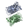
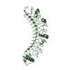
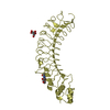
- PDB-3ai5: Crystal structure of yeast enhanced green fluorescent protein-ubi... -
+
Open data
ID or keywords:
Loading...
-
Basic information
Entry
Database: PDB / ID: 3ai5
Title
Crystal structure of yeast enhanced green fluorescent protein-ubiquitin fusion protein
Components
yeast enhanced green fluorescent protein,Ubiquitin
Keywords
Fluorescent Protein / transcription / green fluorescent protein / ubiquitin / fusion protein
Function / homology
Function and homology information
bioluminescence / generation of precursor metabolites and energy / structural constituent of ribosome / ribosome / translation / ribonucleoprotein complex / nucleus / cytoplasm Similarity search - Function
Green Fluorescent Protein / Green fluorescent protein / Ribosomal L40e family / Ribosomal protein L40e / Ribosomal protein L40e superfamily / Green fluorescent protein, GFP / Phosphatidylinositol 3-kinase Catalytic Subunit; Chain A, domain 1 / Green fluorescent protein-related / Green fluorescent protein / Green fluorescent protein ...Green Fluorescent Protein / Green fluorescent protein / Ribosomal L40e family / Ribosomal protein L40e / Ribosomal protein L40e superfamily / Green fluorescent protein, GFP / Phosphatidylinositol 3-kinase Catalytic Subunit; Chain A, domain 1 / Green fluorescent protein-related / Green fluorescent protein / Green fluorescent protein / : / Ubiquitin domain signature. / Ubiquitin conserved site / Ubiquitin-like (UB roll) / Ubiquitin domain / Ubiquitin family / Ubiquitin homologues / Ubiquitin domain profile. / Ubiquitin-like domain / Ubiquitin-like domain superfamily / Roll / Beta Barrel / Mainly Beta / Alpha Beta Similarity search - Domain/homology
Mass: 18.015 Da / Num. of mol.: 299 / Source method: isolated from a natural source / Formula: H2O
Compound details
YEAST ENHANCED GREEN FLUORESCENT PROTEIN - UBIQUITIN FUSION PROTEIN
Has protein modification
Y
Sequence details
THE SEQUENCE OF YEAST ENHANCED GREEN FLUORESCENT PROTEIN HAS BEEN DEPOSITED IN GENBANK WITH ...THE SEQUENCE OF YEAST ENHANCED GREEN FLUORESCENT PROTEIN HAS BEEN DEPOSITED IN GENBANK WITH ACCESSION CODE, ABI82055. residues -2,-1,0 are expression tags, and residue 68 CR2 is CHROMOPHORE (GLY-TYR-GLY).
-
Experimental details
-
Experiment
Experiment
Method: X-RAY DIFFRACTION / Number of used crystals: 1
-
Sample preparation
Crystal
Density Matthews: 2.34 Å3/Da / Density % sol: 47.41 %
Crystal grow
Temperature: 293 K / Method: vapor diffusion, sitting drop / pH: 8 Details: 1% Tryptone, 20% PEG 3350, pH 8.0, VAPOR DIFFUSION, SITTING DROP, temperature 293.0K
In the structure databanks used in Yorodumi, some data are registered as the other names, "COVID-19 virus" and "2019-nCoV". Here are the details of the virus and the list of structure data.
Jan 31, 2019. EMDB accession codes are about to change! (news from PDBe EMDB page)
EMDB accession codes are about to change! (news from PDBe EMDB page)
The allocation of 4 digits for EMDB accession codes will soon come to an end. Whilst these codes will remain in use, new EMDB accession codes will include an additional digit and will expand incrementally as the available range of codes is exhausted. The current 4-digit format prefixed with “EMD-” (i.e. EMD-XXXX) will advance to a 5-digit format (i.e. EMD-XXXXX), and so on. It is currently estimated that the 4-digit codes will be depleted around Spring 2019, at which point the 5-digit format will come into force.
The EM Navigator/Yorodumi systems omit the EMD- prefix.
Related info.:Q: What is EMD? / ID/Accession-code notation in Yorodumi/EM Navigator
Yorodumi is a browser for structure data from EMDB, PDB, SASBDB, etc.
This page is also the successor to EM Navigator detail page, and also detail information page/front-end page for Omokage search.
The word "yorodu" (or yorozu) is an old Japanese word meaning "ten thousand". "mi" (miru) is to see.
Related info.:EMDB / PDB / SASBDB / Comparison of 3 databanks / Yorodumi Search / Aug 31, 2016. New EM Navigator & Yorodumi / Yorodumi Papers / Jmol/JSmol / Function and homology information / Changes in new EM Navigator and Yorodumi
 Movie
Movie Controller

 Yorodumi
 Open data
 Basic information
 Components
 Keywords
 Function and homology information
 Aequorea victoria (jellyfish)
 X-RAY DIFFRACTION /
  Authors
 Citation
 Structure visualization
 Downloads & links
 Other downloads
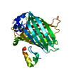


 PDBj
PDBj


 Assembly
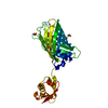


 Mass: 62.068 Da / Num. of mol.: 5 / Source method: obtained synthetically / Formula: C2H6O2
 Mass: 18.015 Da / Num. of mol.: 299 / Source method: isolated from a natural source / Formula: H2O
 Sample preparation
 / Beamline: AR-NW12A / Wavelength: 1 Å
 Processing