Translesion synthesis by POLI / Termination of translesion DNA synthesis / cellular response to UV-C / translesion synthesis / serine-type endopeptidase inhibitor activity / DNA replication / damaged DNA binding / DNA-directed DNA polymerase / DNA-directed DNA polymerase activity / extracellular space ...Translesion synthesis by POLI / Termination of translesion DNA synthesis / cellular response to UV-C / translesion synthesis / serine-type endopeptidase inhibitor activity / DNA replication / damaged DNA binding / DNA-directed DNA polymerase / DNA-directed DNA polymerase activity / extracellular space / nucleus / metal ion binding Similarity search - Function
DNA polymerase type-Y, HhH motif / IMS family HHH motif / DNA polymerase, Y-family, little finger domain / impB/mucB/samB family C-terminal domain / UmuC domain / DNA polymerase, Y-family, little finger domain superfamily / impB/mucB/samB family / UmuC domain profile. / Green Fluorescent Protein / Green fluorescent protein ...DNA polymerase type-Y, HhH motif / IMS family HHH motif / DNA polymerase, Y-family, little finger domain / impB/mucB/samB family C-terminal domain / UmuC domain / DNA polymerase, Y-family, little finger domain superfamily / impB/mucB/samB family / UmuC domain profile. / Green Fluorescent Protein / Green fluorescent protein / Green fluorescent protein / Serpin, conserved site / Serpins signature. / Serpin superfamily, domain 2 / Serpin family / Serpin domain / Serpin superfamily / Serpin superfamily, domain 1 / Serpin (serine protease inhibitor) / SERine Proteinase INhibitors / Reverse transcriptase/Diguanylate cyclase domain / DNA/RNA polymerase superfamily / Beta Barrel / Mainly Beta Similarity search - Domain/homology
Mass: 18.015 Da / Num. of mol.: 296 / Source method: isolated from a natural source / Formula: H2O
Compound details
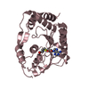
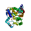
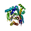
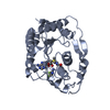
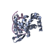
YEAST ENHANCED GREEN FLUORESCENT PROTEIN - MOUSE POLYMERASE IOTA UBIQUITIN BINDING MOTIF FUSION PROTEIN
Sequence details
THE SEQUENCE OF YEAST ENHANCED GREEN FLUORESCENT PROTEIN HAS BEEN DEPOSITED IN GENBANK WITH ...THE SEQUENCE OF YEAST ENHANCED GREEN FLUORESCENT PROTEIN HAS BEEN DEPOSITED IN GENBANK WITH ACCESSION CODE, ABI82055. residues -2,-1,0 are expression tags, and residue 68 CR2 is CHROMOPHORE (GLY-TYR-GLY).
-
Experimental details
-
Experiment
Experiment
Method: X-RAY DIFFRACTION / Number of used crystals: 1
-
Sample preparation
Crystal
Density Matthews: 2.49 Å3/Da / Density % sol: 50.62 %
Crystal grow
Temperature: 293 K / Method: vapor diffusion, sitting drop / pH: 7.5 Details: 0.1M HEPES, 2.0M Ammonium sulfate, pH 7.5, VAPOR DIFFUSION, SITTING DROP, temperature 293.0K
In the structure databanks used in Yorodumi, some data are registered as the other names, "COVID-19 virus" and "2019-nCoV". Here are the details of the virus and the list of structure data.
Jan 31, 2019. EMDB accession codes are about to change! (news from PDBe EMDB page)
EMDB accession codes are about to change! (news from PDBe EMDB page)
The allocation of 4 digits for EMDB accession codes will soon come to an end. Whilst these codes will remain in use, new EMDB accession codes will include an additional digit and will expand incrementally as the available range of codes is exhausted. The current 4-digit format prefixed with “EMD-” (i.e. EMD-XXXX) will advance to a 5-digit format (i.e. EMD-XXXXX), and so on. It is currently estimated that the 4-digit codes will be depleted around Spring 2019, at which point the 5-digit format will come into force.
The EM Navigator/Yorodumi systems omit the EMD- prefix.
Related info.:Q: What is EMD? / ID/Accession-code notation in Yorodumi/EM Navigator
Yorodumi is a browser for structure data from EMDB, PDB, SASBDB, etc.
This page is also the successor to EM Navigator detail page, and also detail information page/front-end page for Omokage search.
The word "yorodu" (or yorozu) is an old Japanese word meaning "ten thousand". "mi" (miru) is to see.
Related info.:EMDB / PDB / SASBDB / Comparison of 3 databanks / Yorodumi Search / Aug 31, 2016. New EM Navigator & Yorodumi / Yorodumi Papers / Jmol/JSmol / Function and homology information / Changes in new EM Navigator and Yorodumi
 Movie
Movie Controller

 Yorodumi
 Open data
 Basic information
 Components
 Keywords
 Function and homology information
 Aequorea victoria (jellyfish)
 X-RAY DIFFRACTION /
  Authors
 Citation
 Structure visualization
 Downloads & links
 Other downloads
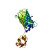


 PDBj
PDBj


 Assembly
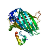


 Mass: 96.063 Da / Num. of mol.: 1 / Source method: obtained synthetically / Formula: SO4
 Mass: 18.015 Da / Num. of mol.: 296 / Source method: isolated from a natural source / Formula: H2O
 Sample preparation
 / Beamline: AR-NE3A / Wavelength: 1 Å
 Processing