 Movie
Movie Controller

[English] 日本語
 Yorodumi
- PDB-3ahf: Phosphoketolase from Bifidobacterium Breve complexed with inorgan... -
+ Open data

- Basic information
| Entry | Database: PDB / ID: 3ahf | ||||||
|---|---|---|---|---|---|---|---|
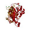
| Title | Phosphoketolase from Bifidobacterium Breve complexed with inorganic phosphate | ||||||
 Components Components | Xylulose 5-phosphate/fructose 6-phosphate phosphoketolase | ||||||
 Keywords Keywords | LYASE / THIAMINE DIPHOSPHATE-DEPENDENT ENZYME / ALPHA-BETA FOLD / HOMODIMER / INORGANIC PHOSPHATE | ||||||
| Function / homology |  Function and homology information Function and homology informationLyases; Carbon-carbon lyases; Aldehyde-lyases / aldehyde-lyase activity / carbohydrate metabolic process / magnesium ion binding Similarity search - Function | ||||||
| Biological species |  Bifidobacterium breve (bacteria) Bifidobacterium breve (bacteria) | ||||||
| Method |  X-RAY DIFFRACTION / SYNCHROTRON / MOLECULAR REPLACEMENT / Resolution: 2.3 Å X-RAY DIFFRACTION / SYNCHROTRON / MOLECULAR REPLACEMENT / Resolution: 2.3 Å | ||||||
 Authors Authors | Suzuki, R. / Katayama, T. / Kim, B.-J. / Wakagi, T. / Shoun, H. / Ashida, H. / Yamamoto, K. / Fushinobu, S. | ||||||
 Citation Citation | Journal: J.Biol.Chem. / Year: 2010 Title: Crystal Structures of phosphoketolase: thiamine diphosphate-dependent dehydration mechanism Authors: Suzuki, R. / Katayama, T. / Kim, B.-J. / Wakagi, T. / Shoun, H. / Ashida, H. / Yamamoto, K. / Fushinobu, S. #1: Journal: Acta Crystallogr.,Sect.F / Year: 2010 Title: Overexpression, crystallization and preliminary X-ray analysis of xylulose-5-phosphate/fructose-6-phosphate phosphoketolase from Bifidobacterium breve Authors: Suzuki, R. / Kim, B.-J. / Shibata, T. / Iwamoto, Y. / Katayama, T. / Ashida, H. / Wakagi, T. / Shoun, H. / Fushinobu, S. / Yamamoto, K. | ||||||
| History |
|
- Structure visualization
| Structure viewer | Molecule: MolmilJmol/JSmol |
|---|
- Downloads & links
-Download
| PDBx/mmCIF format | 3ahf.cif.gz | 188.2 KB | Display | PDBx/mmCIF format |
|---|---|---|---|---|
| PDB format | pdb3ahf.ent.gz | 144.6 KB | Display | PDB format |
| PDBx/mmJSON format | 3ahf.json.gz | Tree view | PDBx/mmJSON format | |
| Others |  Other downloads Other downloads |
-Validation report
| Arichive directory | https://data.pdbj.org/pub/pdb/validation_reports/ah/3ahfftp://data.pdbj.org/pub/pdb/validation_reports/ah/3ahf | HTTPS FTP |
|---|
-Related structure data
| Related structure data |  3ahcSC  3ahdC  3aheC  3ahgC  3ahhC  3ahiC  3ahjC S: Starting model for refinement C: citing same article ( |
|---|---|
| Similar structure data |


-Links
 PDBj
PDBj


- Assembly
| Deposited unit | 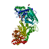
| ||||||||
|---|---|---|---|---|---|---|---|---|---|
| 1 | 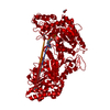
| ||||||||
| Unit cell |
|
-Components
-Protein , 1 types, 1 molecules A
| #1: Protein | Mass: 94962.891 Da / Num. of mol.: 1 Source method: isolated from a genetically manipulated source Source: (gene. exp.) Bifidobacterium breve (bacteria) / Strain: 203 / Gene: xfp / Plasmid: pET28b / Production host: References: UniProt: D6PAH1, fructose-6-phosphate phosphoketolase |
|---|
-Non-polymers , 5 types, 524 molecules 


| #2: Chemical | ChemComp-TPP /  Mass: 425.314 Da / Num. of mol.: 1 / Source method: obtained synthetically / Formula: C12H19N4O7P2S Mass: 425.314 Da / Num. of mol.: 1 / Source method: obtained synthetically / Formula: C12H19N4O7P2S | ||
|---|---|---|---|
| #3: Chemical | ChemComp-MG /  Mass: 24.305 Da / Num. of mol.: 1 / Source method: obtained synthetically / Formula: Mg Mass: 24.305 Da / Num. of mol.: 1 / Source method: obtained synthetically / Formula: Mg | ||
| #4: Chemical | ChemComp-PO4 /  Mass: 94.971 Da / Num. of mol.: 1 / Source method: obtained synthetically / Formula: PO4 Mass: 94.971 Da / Num. of mol.: 1 / Source method: obtained synthetically / Formula: PO4 | ||
| #5: Chemical |  Mass: 92.094 Da / Num. of mol.: 3 / Source method: obtained synthetically / Formula: C3H8O3 Mass: 92.094 Da / Num. of mol.: 3 / Source method: obtained synthetically / Formula: C3H8O3#6: Water | ChemComp-HOH / | Mass: 18.015 Da / Num. of mol.: 518 / Source method: isolated from a natural source / Formula: H2O |
-Experimental details
-Experiment
| Experiment | Method: X-RAY DIFFRACTION / Number of used crystals: 1 |
|---|
- Sample preparation
| Crystal | Density Matthews: 3.33 Å3/Da / Density % sol: 63.07 % |
|---|---|
| Crystal grow | Temperature: 293 K / Method: vapor diffusion, sitting drop / pH: 9 Details: 24%(v/v) PEG 6000, 0.1M BICINE buffer, pH 9.0, VAPOR DIFFUSION, SITTING DROP, temperature 293K |
-Data collection
| Diffraction | Mean temperature: 100 K |
|---|---|
| Diffraction source | Source: SYNCHROTRON / Site: Photon Factory  / Beamline: BL-6A / Wavelength: 1 Å / Beamline: BL-6A / Wavelength: 1 Å |
| Detector | Type: ADSC QUANTUM 4r / Detector: CCD / Date: Oct 30, 2008 / Details: mirrors |
| Radiation | Monochromator: Si(111) monochromator / Protocol: SINGLE WAVELENGTH / Monochromatic (M) / Laue (L): M / Scattering type: x-ray |
| Radiation wavelength | Wavelength: 1 Å / Relative weight: 1 |
| Reflection | Resolution: 2.3→50 Å / Num. all: 55669 / Num. obs: 55630 / % possible obs: 100 % / Observed criterion σ(F): 0 / Observed criterion σ(I): 0 / Redundancy: 11.4 % / Biso Wilson estimate: 28.3 Å2 / Rmerge(I) obs: 0.108 / Net I/σ(I): 24.9 |
| Reflection shell | Resolution: 2.3→2.38 Å / Redundancy: 10.4 % / Rmerge(I) obs: 0.355 / Mean I/σ(I) obs: 4.7 / Num. unique all: 5490 / % possible all: 100 |
- Processing
| Software |
| ||||||||||||||||||||||||||||||||||||||||||||||||||||||||||||||||||||||||||||||||||||||||||||||||||||||||||||||||||||||||||||||||||||||||||||||||||||||||||||||||||||||||||
|---|---|---|---|---|---|---|---|---|---|---|---|---|---|---|---|---|---|---|---|---|---|---|---|---|---|---|---|---|---|---|---|---|---|---|---|---|---|---|---|---|---|---|---|---|---|---|---|---|---|---|---|---|---|---|---|---|---|---|---|---|---|---|---|---|---|---|---|---|---|---|---|---|---|---|---|---|---|---|---|---|---|---|---|---|---|---|---|---|---|---|---|---|---|---|---|---|---|---|---|---|---|---|---|---|---|---|---|---|---|---|---|---|---|---|---|---|---|---|---|---|---|---|---|---|---|---|---|---|---|---|---|---|---|---|---|---|---|---|---|---|---|---|---|---|---|---|---|---|---|---|---|---|---|---|---|---|---|---|---|---|---|---|---|---|---|---|---|---|---|---|---|
| Refinement | Method to determine structure: MOLECULAR REPLACEMENT Starting model: PDB ENTRY 3AHC Resolution: 2.3→40.88 Å / Cor.coef. Fo:Fc: 0.956 / Cor.coef. Fo:Fc free: 0.93 / SU B: 5.556 / SU ML: 0.134 / Cross valid method: THROUGHOUT / ESU R: 0.219 / ESU R Free: 0.192 / Stereochemistry target values: MAXIMUM LIKELIHOOD / Details: HYDROGENS HAVE BEEN ADDED IN THE RIDING POSITIONS
| ||||||||||||||||||||||||||||||||||||||||||||||||||||||||||||||||||||||||||||||||||||||||||||||||||||||||||||||||||||||||||||||||||||||||||||||||||||||||||||||||||||||||||
| Solvent computation | Ion probe radii: 0.8 Å / Shrinkage radii: 0.8 Å / VDW probe radii: 1.4 Å / Solvent model: BABINET MODEL WITH MASK | ||||||||||||||||||||||||||||||||||||||||||||||||||||||||||||||||||||||||||||||||||||||||||||||||||||||||||||||||||||||||||||||||||||||||||||||||||||||||||||||||||||||||||
| Displacement parameters | Biso mean: 28.516 Å2
| ||||||||||||||||||||||||||||||||||||||||||||||||||||||||||||||||||||||||||||||||||||||||||||||||||||||||||||||||||||||||||||||||||||||||||||||||||||||||||||||||||||||||||
| Refine analyze |
| ||||||||||||||||||||||||||||||||||||||||||||||||||||||||||||||||||||||||||||||||||||||||||||||||||||||||||||||||||||||||||||||||||||||||||||||||||||||||||||||||||||||||||
| Refinement step | Cycle: LAST / Resolution: 2.3→40.88 Å
| ||||||||||||||||||||||||||||||||||||||||||||||||||||||||||||||||||||||||||||||||||||||||||||||||||||||||||||||||||||||||||||||||||||||||||||||||||||||||||||||||||||||||||
| Refine LS restraints |
| ||||||||||||||||||||||||||||||||||||||||||||||||||||||||||||||||||||||||||||||||||||||||||||||||||||||||||||||||||||||||||||||||||||||||||||||||||||||||||||||||||||||||||
| LS refinement shell | Resolution: 2.299→2.359 Å / Total num. of bins used: 20
|
