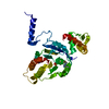
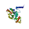
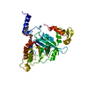
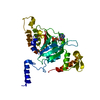
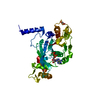
 Movie
Movie Controller


+ Open data

- Basic information
| Entry | Database: PDB / ID: 2zrd | ||||||
|---|---|---|---|---|---|---|---|
| Title | MsRecA Q196N ADP form IV | ||||||
 Components Components | Protein recA | ||||||
 Keywords Keywords | HYDROLASE / recombination / RecA mutants / DNA-REPAIR / ATP-binding / Cytoplasm / DNA damage / DNA recombination / DNA repair / DNA-binding / Nucleotide-binding / SOS response | ||||||
| Function / homology |  Function and homology information Function and homology informationSOS response / ATP-dependent DNA damage sensor activity / single-stranded DNA binding / DNA recombination / damaged DNA binding / DNA repair / ATP hydrolysis activity / ATP binding / cytosol Similarity search - Function | ||||||
| Biological species |  Mycobacterium smegmatis str. MC2 155 (bacteria) Mycobacterium smegmatis str. MC2 155 (bacteria) | ||||||
| Method |  X-RAY DIFFRACTION / MOLECULAR REPLACEMENT / Resolution: 3.1 Å X-RAY DIFFRACTION / MOLECULAR REPLACEMENT / Resolution: 3.1 Å | ||||||
 Authors Authors | Prabu, J.R. / Manjunath, G.P. / Chandra, N.R. / Muniyappa, K. / Vijayan, M. | ||||||
 Citation Citation | Journal: Acta Crystallogr.,Sect.D / Year: 2008 Title: Functionally important movements in RecA molecules and filaments: studies involving mutation and environmental changes Authors: Prabu, J.R. / Manjunath, G.P. / Chandra, N.R. / Muniyappa, K. / Vijayan, M. | ||||||
| History |
|
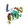
- Structure visualization
| Structure viewer | Molecule: MolmilJmol/JSmol |
|---|
- Downloads & links
-Download
| PDBx/mmCIF format | 2zrd.cif.gz | 69 KB | Display | PDBx/mmCIF format |
|---|---|---|---|---|
| PDB format | pdb2zrd.ent.gz | 50.3 KB | Display | PDB format |
| PDBx/mmJSON format | 2zrd.json.gz | Tree view | PDBx/mmJSON format | |
| Others |  Other downloads Other downloads |
-Validation report
| Arichive directory | https://data.pdbj.org/pub/pdb/validation_reports/zr/2zrdftp://data.pdbj.org/pub/pdb/validation_reports/zr/2zrd | HTTPS FTP |
|---|
-Related structure data
| Related structure data |  2zr0C  2zr7C  2zr9C 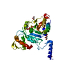 2zraC  2zrbC  2zrcC 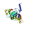 2zreC  2zrfC  2zrgC  2zrhC  2zriC 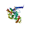 2zrjC  2zrkC  2zrlC  2zrmC  2zrnC  2zroC  2zrpC 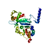 1ubcS S: Starting model for refinement C: citing same article ( |
|---|---|
| Similar structure data |


-Links
 PDBj
PDBj
- Assembly
| Deposited unit | 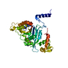
| ||||||||
|---|---|---|---|---|---|---|---|---|---|
| 1 |
| ||||||||
| Unit cell |
|
-Components
| #1: Protein | Mass: 37330.465 Da / Num. of mol.: 1 / Mutation: Q196N Source method: isolated from a genetically manipulated source Source: (gene. exp.) Mycobacterium smegmatis str. MC2 155 (bacteria)Plasmid: PTHIOA / Production host: |
|---|---|
| #2: Chemical | ChemComp-ADP /   Mass: 427.201 Da / Num. of mol.: 1 / Source method: obtained synthetically / Formula: C10H15N5O10P2 / Comment: ADP, energy-carrying molecule*YM Mass: 427.201 Da / Num. of mol.: 1 / Source method: obtained synthetically / Formula: C10H15N5O10P2 / Comment: ADP, energy-carrying molecule*YM |
| #3: Water | ChemComp-HOH /  Mass: 18.015 Da / Num. of mol.: 34 / Source method: isolated from a natural source / Formula: H2O Mass: 18.015 Da / Num. of mol.: 34 / Source method: isolated from a natural source / Formula: H2O |
-Experimental details
-Experiment
| Experiment | Method: X-RAY DIFFRACTION / Number of used crystals: 1 |
|---|
- Sample preparation
| Crystal | Density Matthews: 3.07 Å3/Da / Density % sol: 59.91 % |
|---|---|
| Crystal grow | Temperature: 296 K / Method: vapor diffusion, hanging drop / pH: 6.9 Details: 80mM citrate/phosphate buffer(pH 6.9), 80mM NaCl, 40mM ammonium acetate, 20mM sodium citrate, 6% poly-ethylene glycol 3350, 30% poly-ethylene glycol 3350, 200mM ammonium acetate in sodium ...Details: 80mM citrate/phosphate buffer(pH 6.9), 80mM NaCl, 40mM ammonium acetate, 20mM sodium citrate, 6% poly-ethylene glycol 3350, 30% poly-ethylene glycol 3350, 200mM ammonium acetate in sodium citrate buffer(PH 5.8), VAPOR DIFFUSION, HANGING DROP, temperature 296K |
-Data collection
| Diffraction | Mean temperature: 100 K |
|---|---|
| Diffraction source | Source: ROTATING ANODE / Type: RIGAKU RU200 / Wavelength: 1.54 Å |
| Detector | Type: MAR scanner 345 mm plate / Detector: IMAGE PLATE / Date: Jun 15, 2007 / Details: Mirrors |
| Radiation | Monochromator: Single crystal, cylindrically bent, Si(220) / Protocol: SINGLE WAVELENGTH / Monochromatic (M) / Laue (L): M / Scattering type: x-ray |
| Radiation wavelength | Wavelength: 1.54 Å / Relative weight: 1 |
| Reflection | Resolution: 3.1→30 Å / Num. all: 8273 / Num. obs: 8220 / % possible obs: 99.3 % / Observed criterion σ(I): 0 / Redundancy: 4.3 % / Rmerge(I) obs: 0.107 / Net I/σ(I): 13.6 |
| Reflection shell | Resolution: 3.1→3.27 Å / Redundancy: 3.1 % / Rmerge(I) obs: 0.397 / Mean I/σ(I) obs: 2.6 / Num. unique all: 1172 / % possible all: 98 |
- Processing
| Software |
| ||||||||||||||||||||||||||||||||||||||||||||||||||||||||||||
|---|---|---|---|---|---|---|---|---|---|---|---|---|---|---|---|---|---|---|---|---|---|---|---|---|---|---|---|---|---|---|---|---|---|---|---|---|---|---|---|---|---|---|---|---|---|---|---|---|---|---|---|---|---|---|---|---|---|---|---|---|---|
| Refinement | Method to determine structure: MOLECULAR REPLACEMENT Starting model: 1UBC Resolution: 3.1→29.49 Å / Rfactor Rfree error: 0.009 / Data cutoff high absF: 1071145.39 / Data cutoff low absF: 0 / Isotropic thermal model: GROUP / Cross valid method: THROUGHOUT / σ(F): 0 / Stereochemistry target values: Engh & Huber / Details: BULK SOLVENT MODEL USED
| ||||||||||||||||||||||||||||||||||||||||||||||||||||||||||||
| Solvent computation | Solvent model: FLAT MODEL / Bsol: 46.967 Å2 / ksol: 0.35 e/Å3 | ||||||||||||||||||||||||||||||||||||||||||||||||||||||||||||
| Displacement parameters | Biso mean: 46.4 Å2
| ||||||||||||||||||||||||||||||||||||||||||||||||||||||||||||
| Refine analyze |
| ||||||||||||||||||||||||||||||||||||||||||||||||||||||||||||
| Refinement step | Cycle: LAST / Resolution: 3.1→29.49 Å
| ||||||||||||||||||||||||||||||||||||||||||||||||||||||||||||
| Refine LS restraints |
| ||||||||||||||||||||||||||||||||||||||||||||||||||||||||||||
| LS refinement shell | Resolution: 3.1→3.29 Å / Rfactor Rfree error: 0.03 / Total num. of bins used: 6
| ||||||||||||||||||||||||||||||||||||||||||||||||||||||||||||
| Xplor file |
|
