 Movie
Movie Controller

[English] 日本語
 Yorodumi
- PDB-2yzu: Crystal structure of oxidized thioredoxin from Thermus thermophil... -
+ Open data

- Basic information
| Entry | Database: PDB / ID: 2yzu | ||||||
|---|---|---|---|---|---|---|---|
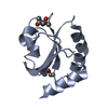
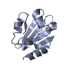
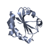
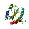
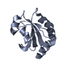
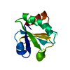
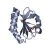
| Title | Crystal structure of oxidized thioredoxin from Thermus thermophilus HB8 | ||||||
 Components Components | Thioredoxin | ||||||
 Keywords Keywords | ELECTRON TRANSPORT / Redox protein / Structural Genomics / NPPSFA / National Project on Protein Structural and Functional Analyses / RIKEN Structural Genomics/Proteomics Initiative / RSGI | ||||||
| Function / homology |  Function and homology information Function and homology informationglycerol ether metabolic process / protein-disulfide reductase activity / cell redox homeostasis Similarity search - Function | ||||||
| Biological species |   Thermus thermophilus (bacteria) Thermus thermophilus (bacteria) | ||||||
| Method |  X-RAY DIFFRACTION / SYNCHROTRON / MOLECULAR REPLACEMENT / Resolution: 1.9 Å X-RAY DIFFRACTION / SYNCHROTRON / MOLECULAR REPLACEMENT / Resolution: 1.9 Å | ||||||
 Authors Authors | Ebihara, A. / Yanai, H. / Yokoyama, S. / Kuramitsu, S. / RIKEN Structural Genomics/Proteomics Initiative (RSGI) | ||||||
 Citation Citation | Journal: To be Published Title: Crystal structure of oxidized thioredoxin from Thermus thermophilus HB8 Authors: Ebihara, A. / Yanai, H. / Yokoyama, S. / Kuramitsu, S. | ||||||
| History |
|
- Structure visualization
| Structure viewer | Molecule: MolmilJmol/JSmol |
|---|
- Downloads & links
-Download
| PDBx/mmCIF format | 2yzu.cif.gz | 34 KB | Display | PDBx/mmCIF format |
|---|---|---|---|---|
| PDB format | pdb2yzu.ent.gz | 22 KB | Display | PDB format |
| PDBx/mmJSON format | 2yzu.json.gz | Tree view | PDBx/mmJSON format | |
| Others |  Other downloads Other downloads |
-Validation report
| Summary document | 2yzu_validation.pdf.gz | 425.2 KB | Display | wwPDB validaton report |
|---|---|---|---|---|
| Full document | 2yzu_full_validation.pdf.gz | 427.4 KB | Display | |
| Data in XML | 2yzu_validation.xml.gz | 6.6 KB | Display | |
| Data in CIF | 2yzu_validation.cif.gz | 8 KB | Display | |
| Arichive directory | https://data.pdbj.org/pub/pdb/validation_reports/yz/2yzuftp://data.pdbj.org/pub/pdb/validation_reports/yz/2yzu | HTTPS FTP |
-Related structure data
| Related structure data |  2cvkS S: Starting model for refinement |
|---|---|
| Similar structure data | |
| Other databases |


-Links
 PDBj
PDBj

- Assembly
| Deposited unit | 
| ||||||||
|---|---|---|---|---|---|---|---|---|---|
| 1 |
| ||||||||
| Unit cell |
|
-Components
| #1: Protein | Mass: 12486.024 Da / Num. of mol.: 1 / Fragment: Residues 2-110 Source method: isolated from a genetically manipulated source Source: (gene. exp.) Thermus thermophilus (bacteria) / Strain: HB8 / Plasmid: pET-11a / Production host: |
|---|---|
| #2: Water | ChemComp-HOH /  Mass: 18.015 Da / Num. of mol.: 33 / Source method: isolated from a natural source / Formula: H2O Mass: 18.015 Da / Num. of mol.: 33 / Source method: isolated from a natural source / Formula: H2O |
-Experimental details
-Experiment
| Experiment | Method: X-RAY DIFFRACTION / Number of used crystals: 1 |
|---|
- Sample preparation
| Crystal | Density Matthews: 2.04 Å3/Da / Density % sol: 39.76 % |
|---|---|
| Crystal grow | Temperature: 293 K / Method: vapor diffusion, sitting drop / pH: 6.8 Details: 4.3M Sodium chloride, 0.1M HEPES, pH 6.8, VAPOR DIFFUSION, SITTING DROP, temperature 293K |
-Data collection
| Diffraction | Mean temperature: 100 K |
|---|---|
| Diffraction source | Source: SYNCHROTRON / Site: SPring-8  / Beamline: BL26B1 / Wavelength: 1 Å / Beamline: BL26B1 / Wavelength: 1 Å |
| Detector | Type: RIGAKU JUPITER 210 / Detector: CCD / Date: Jul 19, 2005 |
| Radiation | Monochromator: SI Double-Crystal / Protocol: SINGLE WAVELENGTH / Monochromatic (M) / Laue (L): M / Scattering type: x-ray |
| Radiation wavelength | Wavelength: 1 Å / Relative weight: 1 |
| Reflection | Resolution: 1.9→50 Å / Num. obs: 8643 / % possible obs: 99.7 % / Redundancy: 13.3 % / Biso Wilson estimate: 26.7 Å2 / Rmerge(I) obs: 0.034 / Net I/σ(I): 60.5 |
| Reflection shell | Resolution: 1.9→1.97 Å / Redundancy: 13.3 % / Rmerge(I) obs: 0.16 / Mean I/σ(I) obs: 17.2 / % possible all: 100 |
- Processing
| Software |
| ||||||||||||||||||||||||||||||||||||||||||||||||||||||||||||||||||||||||||||||||
|---|---|---|---|---|---|---|---|---|---|---|---|---|---|---|---|---|---|---|---|---|---|---|---|---|---|---|---|---|---|---|---|---|---|---|---|---|---|---|---|---|---|---|---|---|---|---|---|---|---|---|---|---|---|---|---|---|---|---|---|---|---|---|---|---|---|---|---|---|---|---|---|---|---|---|---|---|---|---|---|---|---|
| Refinement | Method to determine structure: MOLECULAR REPLACEMENT Starting model: PDB ENTRY 2CVK Resolution: 1.9→20.9 Å / Rfactor Rfree error: 0.009 / Data cutoff high absF: 1027395.22 / Data cutoff low absF: 0 / Isotropic thermal model: RESTRAINED / Cross valid method: THROUGHOUT / σ(F): 0 / Stereochemistry target values: Engh & Huber
| ||||||||||||||||||||||||||||||||||||||||||||||||||||||||||||||||||||||||||||||||
| Solvent computation | Solvent model: FLAT MODEL / Bsol: 50.115 Å2 / ksol: 0.398074 e/Å3 | ||||||||||||||||||||||||||||||||||||||||||||||||||||||||||||||||||||||||||||||||
| Displacement parameters | Biso mean: 32 Å2
| ||||||||||||||||||||||||||||||||||||||||||||||||||||||||||||||||||||||||||||||||
| Refine analyze |
| ||||||||||||||||||||||||||||||||||||||||||||||||||||||||||||||||||||||||||||||||
| Refinement step | Cycle: LAST / Resolution: 1.9→20.9 Å
| ||||||||||||||||||||||||||||||||||||||||||||||||||||||||||||||||||||||||||||||||
| Refine LS restraints |
| ||||||||||||||||||||||||||||||||||||||||||||||||||||||||||||||||||||||||||||||||
| LS refinement shell | Resolution: 1.9→2.02 Å / Rfactor Rfree error: 0.032 / Total num. of bins used: 6
| ||||||||||||||||||||||||||||||||||||||||||||||||||||||||||||||||||||||||||||||||
| Xplor file |
|
