 Movie
Movie Controller

[English] 日本語
 Yorodumi
- PDB-2yqc: Crystal Structure of uridine-diphospho-N-acetylglucosamine pyroph... -
+ Open data

- Basic information
| Entry | Database: PDB / ID: 2yqc | ||||||
|---|---|---|---|---|---|---|---|
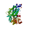
| Title | Crystal Structure of uridine-diphospho-N-acetylglucosamine pyrophosphorylase from Candida albicans, in the apo-like form | ||||||
 Components Components | UDP-N-acetylglucosamine pyrophosphorylase | ||||||
 Keywords Keywords | TRANSFERASE / Pyrophosphorylase / N-acetylglucosamine / Uridine-diphospho-N-acetylglucosamine / N-acetylglucosamine-1-phosphate / Candida albicans | ||||||
| Function / homology |  Function and homology information Function and homology informationUDP-N-acetylgalactosamine metabolic process / UDP-N-acetylgalactosamine diphosphorylase activity / UDP-N-acetylglucosamine diphosphorylase / UDP-N-acetylglucosamine diphosphorylase activity / UDP-N-acetylglucosamine biosynthetic process / cytoplasm Similarity search - Function | ||||||
| Biological species |  Candida albicans (yeast) Candida albicans (yeast) | ||||||
| Method |  X-RAY DIFFRACTION / SYNCHROTRON / MOLECULAR REPLACEMENT / Resolution: 1.9 Å X-RAY DIFFRACTION / SYNCHROTRON / MOLECULAR REPLACEMENT / Resolution: 1.9 Å | ||||||
 Authors Authors | Miki, K. / Maruyama, D. / Nishitani, Y. / Nonaka, T. / Kita, A. | ||||||
 Citation Citation | Journal: J.Biol.Chem. / Year: 2007 Title: Crystal Structure of Uridine-diphospho-N-acetylglucosamine Pyrophosphorylase from Candida albicans and Catalytic Reaction Mechanism Authors: Maruyama, D. / Nishitani, Y. / Nonaka, T. / Kita, A. / Fukami, T.A. / Mio, T. / Yamada-Okabe, H. / Yamada-Okabe, T. / Miki, K. | ||||||
| History |
|
- Structure visualization
| Structure viewer | Molecule: MolmilJmol/JSmol |
|---|
- Downloads & links
-Download
| PDBx/mmCIF format | 2yqc.cif.gz | 119.9 KB | Display | PDBx/mmCIF format |
|---|---|---|---|---|
| PDB format | pdb2yqc.ent.gz | 90.2 KB | Display | PDB format |
| PDBx/mmJSON format | 2yqc.json.gz | Tree view | PDBx/mmJSON format | |
| Others |  Other downloads Other downloads |
-Validation report
| Arichive directory | https://data.pdbj.org/pub/pdb/validation_reports/yq/2yqcftp://data.pdbj.org/pub/pdb/validation_reports/yq/2yqc | HTTPS FTP |
|---|
-Related structure data
| Related structure data | 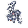 2yqhC 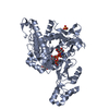 2yqjC 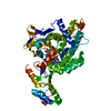 2yqsC 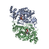 1jvdS C: citing same article ( S: Starting model for refinement |
|---|---|
| Similar structure data |


-Links
 PDBj
PDBj

- Assembly
| Deposited unit | 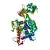
| ||||||||
|---|---|---|---|---|---|---|---|---|---|
| 1 |
| ||||||||
| Unit cell |
|
-Components
| #1: Protein | Mass: 54727.832 Da / Num. of mol.: 1 / Mutation: S216L Source method: isolated from a genetically manipulated source Source: (gene. exp.) Candida albicans (yeast) / Plasmid: pGEX-2T / Production host:  References: UniProt: O74933, UDP-N-acetylglucosamine diphosphorylase |
|---|---|
| #2: Chemical | ChemComp-MG /   Mass: 24.305 Da / Num. of mol.: 1 / Source method: obtained synthetically / Formula: Mg Mass: 24.305 Da / Num. of mol.: 1 / Source method: obtained synthetically / Formula: Mg |
| #3: Chemical | ChemComp-GOL /   Mass: 92.094 Da / Num. of mol.: 1 / Source method: obtained synthetically / Formula: C3H8O3 Mass: 92.094 Da / Num. of mol.: 1 / Source method: obtained synthetically / Formula: C3H8O3 |
| #4: Water | ChemComp-HOH /  Mass: 18.015 Da / Num. of mol.: 436 / Source method: isolated from a natural source / Formula: H2O Mass: 18.015 Da / Num. of mol.: 436 / Source method: isolated from a natural source / Formula: H2O |
-Experimental details
-Experiment
| Experiment | Method: X-RAY DIFFRACTION / Number of used crystals: 1 |
|---|
- Sample preparation
| Crystal | Density Matthews: 2.46 Å3/Da / Density % sol: 50.06 % |
|---|---|
| Crystal grow | Temperature: 293 K / Method: vapor diffusion, sitting drop / pH: 5.5 Details: 30% PEG 6000, 0.1M ammonium sulfate, 5% glycerol, 0.001M N-acetylglucosamine-1-phosphate, 0.1M citrate, pH 5.5, VAPOR DIFFUSION, SITTING DROP, temperature 293.0K |
-Data collection
| Diffraction | Mean temperature: 95 K |
|---|---|
| Diffraction source | Source: SYNCHROTRON / Site: Photon Factory  / Beamline: AR-NW12A / Wavelength: 1 Å / Beamline: AR-NW12A / Wavelength: 1 Å |
| Detector | Type: ADSC QUANTUM 210 / Detector: CCD / Date: Oct 28, 2003 |
| Radiation | Monochromator: Si(111) double crystal monochromator / Protocol: SINGLE WAVELENGTH / Monochromatic (M) / Laue (L): M / Scattering type: x-ray |
| Radiation wavelength | Wavelength: 1 Å / Relative weight: 1 |
| Reflection | Resolution: 1.9→50 Å / Num. obs: 40383 / % possible obs: 90.2 % / Redundancy: 3.5 % / Biso Wilson estimate: 17.8 Å2 / Rsym value: 0.111 / Net I/σ(I): 11.6 |
| Reflection shell | Resolution: 1.9→1.97 Å / Redundancy: 2.5 % / Mean I/σ(I) obs: 2.6 / Num. unique all: 3729 / Rsym value: 0.337 / % possible all: 68.4 |
- Processing
| Software |
| ||||||||||||||||||||||||||||||||||||||||||||||||||||||||||||||||||||||||||||||||
|---|---|---|---|---|---|---|---|---|---|---|---|---|---|---|---|---|---|---|---|---|---|---|---|---|---|---|---|---|---|---|---|---|---|---|---|---|---|---|---|---|---|---|---|---|---|---|---|---|---|---|---|---|---|---|---|---|---|---|---|---|---|---|---|---|---|---|---|---|---|---|---|---|---|---|---|---|---|---|---|---|---|
| Refinement | Method to determine structure: MOLECULAR REPLACEMENT Starting model: PDB ENTRY 1JVD Resolution: 1.9→39.7 Å / Rfactor Rfree error: 0.004 / Data cutoff high absF: 1219636.58 / Data cutoff low absF: 0 / Isotropic thermal model: RESTRAINED / Cross valid method: THROUGHOUT / σ(F): 0 / Stereochemistry target values: Engh & Huber Details: The intensity of the reflection used for the refinement is more than 0 sigma, but that of the unique reflections in the table of the statistics of the X-ray crystallography at the article is ...Details: The intensity of the reflection used for the refinement is more than 0 sigma, but that of the unique reflections in the table of the statistics of the X-ray crystallography at the article is more than 1 sigma. Therefore, the number of reflections in the refinement section is larger than that in the data collection.
| ||||||||||||||||||||||||||||||||||||||||||||||||||||||||||||||||||||||||||||||||
| Solvent computation | Solvent model: FLAT MODEL / Bsol: 51.1034 Å2 / ksol: 0.353506 e/Å3 | ||||||||||||||||||||||||||||||||||||||||||||||||||||||||||||||||||||||||||||||||
| Displacement parameters | Biso mean: 24.3 Å2
| ||||||||||||||||||||||||||||||||||||||||||||||||||||||||||||||||||||||||||||||||
| Refine analyze |
| ||||||||||||||||||||||||||||||||||||||||||||||||||||||||||||||||||||||||||||||||
| Refinement step | Cycle: LAST / Resolution: 1.9→39.7 Å
| ||||||||||||||||||||||||||||||||||||||||||||||||||||||||||||||||||||||||||||||||
| Refine LS restraints |
| ||||||||||||||||||||||||||||||||||||||||||||||||||||||||||||||||||||||||||||||||
| LS refinement shell | Resolution: 1.9→2.02 Å / Rfactor Rfree error: 0.015 / Total num. of bins used: 6
| ||||||||||||||||||||||||||||||||||||||||||||||||||||||||||||||||||||||||||||||||
| Xplor file |
|
