 Movie
Movie Controller

[English] 日本語
 Yorodumi
- PDB-2xvk: crystal structure of alpha-xylosidase (GH31) from Cellvibrio japo... -
+ Open data

- Basic information
| Entry | Database: PDB / ID: 2xvk | ||||||
|---|---|---|---|---|---|---|---|
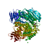
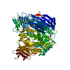
| Title | crystal structure of alpha-xylosidase (GH31) from Cellvibrio japonicus in complex with 5-fluoro-alpha-D-xylopyranosyl fluoride | ||||||
 Components Components | ALPHA-XYLOSIDASE, PUTATIVE, XYL31A | ||||||
 Keywords Keywords | HYDROLASE / GLYCOSYL HYDROLASE FAMILY 31 / (BETA/ALPHA) 8 BARREL | ||||||
| Function / homology |  Function and homology information Function and homology informationHydrolases; Glycosylases; Glycosidases, i.e. enzymes that hydrolyse O- and S-glycosyl compounds / hydrolase activity, hydrolyzing O-glycosyl compounds / carbohydrate binding / carbohydrate metabolic process Similarity search - Function | ||||||
| Biological species |  CELLVIBRIO JAPONICUS (bacteria) CELLVIBRIO JAPONICUS (bacteria) | ||||||
| Method |  X-RAY DIFFRACTION / SYNCHROTRON / MOLECULAR REPLACEMENT / Resolution: 2.503 Å X-RAY DIFFRACTION / SYNCHROTRON / MOLECULAR REPLACEMENT / Resolution: 2.503 Å | ||||||
 Authors Authors | Larsbrink, J. / Izumi, A. / Ibatullin, F. / Nakhai, A. / Gilbert, H.J. / Davies, G.J. / Brumer, H. | ||||||
 Citation Citation | Journal: Biochem.J. / Year: 2011 Title: Structural and Enzymatic Characterisation of a Glycoside Hydrolase Family 31 Alpha-Xylosidase from Cellvibrio Japonicus Involved in Xyloglucan Saccharification. Authors: Larsbrink, J. / Izumi, A. / Ibatullin, F. / Nakhai, A. / Gilbert, H.J. / Davies, G.J. / Brumer, H. | ||||||
| History |
|
- Structure visualization
| Structure viewer | Molecule: MolmilJmol/JSmol |
|---|
- Downloads & links
-Download
| PDBx/mmCIF format | 2xvk.cif.gz | 397.1 KB | Display | PDBx/mmCIF format |
|---|---|---|---|---|
| PDB format | pdb2xvk.ent.gz | 325.2 KB | Display | PDB format |
| PDBx/mmJSON format | 2xvk.json.gz | Tree view | PDBx/mmJSON format | |
| Others |  Other downloads Other downloads |
-Validation report
| Arichive directory | https://data.pdbj.org/pub/pdb/validation_reports/xv/2xvkftp://data.pdbj.org/pub/pdb/validation_reports/xv/2xvk | HTTPS FTP |
|---|
-Related structure data
| Related structure data |  2xvgC  2xvlC 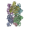 2g3mS C: citing same article ( S: Starting model for refinement |
|---|---|
| Similar structure data |


-Links
 PDBj
PDBj


- Assembly
| Deposited unit | 
| ||||||||
|---|---|---|---|---|---|---|---|---|---|
| 1 |
| ||||||||
| Unit cell |
|
-Components
| #1: Protein | Mass: 115203.914 Da / Num. of mol.: 1 Source method: isolated from a genetically manipulated source Details: THE OD ATOM OF D582 LINKS TO THE C1 ATOM OF FFX. / Source: (gene. exp.) CELLVIBRIO JAPONICUS (bacteria) / Production host: References: UniProt: B3PBD9, Hydrolases; Glycosylases; Glycosidases, i.e. enzymes that hydrolyse O- and S-glycosyl compounds | ||||||||||
|---|---|---|---|---|---|---|---|---|---|---|---|
| #2: Chemical | ChemComp-CL /   Mass: 35.453 Da / Num. of mol.: 4 / Source method: obtained synthetically / Formula: Cl Mass: 35.453 Da / Num. of mol.: 4 / Source method: obtained synthetically / Formula: Cl#3: Chemical | ChemComp-NI / |   Mass: 58.693 Da / Num. of mol.: 1 / Source method: obtained synthetically / Formula: Ni Mass: 58.693 Da / Num. of mol.: 1 / Source method: obtained synthetically / Formula: Ni#4: Sugar | ChemComp-FFX / ( |   Type: D-saccharide / Mass: 170.111 Da / Num. of mol.: 1 / Source method: obtained synthetically / Formula: C5H8F2O4 Type: D-saccharide / Mass: 170.111 Da / Num. of mol.: 1 / Source method: obtained synthetically / Formula: C5H8F2O4#5: Water | ChemComp-HOH / |  Mass: 18.015 Da / Num. of mol.: 83 / Source method: isolated from a natural source / Formula: H2O Mass: 18.015 Da / Num. of mol.: 83 / Source method: isolated from a natural source / Formula: H2OHas protein modification | Y | Nonpolymer details | 5-FLUORO-ALPHA-D-XYLOPYRANOSYL FLUORIDE (FFX): THE FLUORIDE ATOM AT C1 OF THIS LIGAND WAS REMOVED ...5-FLUORO-ALPHA-D-XYLOPYRANO | |
-Experimental details
-Experiment
| Experiment | Method: X-RAY DIFFRACTION / Number of used crystals: 1 |
|---|
- Sample preparation
| Crystal | Density Matthews: 3.5 Å3/Da / Density % sol: 64.81 % / Description: NONE |
|---|---|
| Crystal grow | pH: 7 Details: 25 % PEG MONOMETHYL ETHER 550 (PEG MME 550), 0.1 M BIS-TRIS (PH 7.0) |
-Data collection
| Diffraction | Mean temperature: 100 K |
|---|---|
| Diffraction source | Source: SYNCHROTRON / Site: ESRF  / Beamline: ID29 / Wavelength: 0.9537 / Beamline: ID29 / Wavelength: 0.9537 |
| Detector | Type: ADSC QUANTUM 315r / Detector: CCD / Date: Nov 28, 2009 / Details: MIRRORS |
| Radiation | Monochromator: SI (311) / Protocol: SINGLE WAVELENGTH / Monochromatic (M) / Laue (L): M / Scattering type: x-ray |
| Radiation wavelength | Wavelength: 0.9537 Å / Relative weight: 1 |
| Reflection | Resolution: 2.5→50 Å / Num. obs: 57181 / % possible obs: 99 % / Observed criterion σ(I): -3 / Redundancy: 10.1 % / Rmerge(I) obs: 0.07 / Net I/σ(I): 10.1 |
| Reflection shell | Resolution: 2.5→2.54 Å / Redundancy: 8 % / Rmerge(I) obs: 0.75 / Mean I/σ(I) obs: 2 / % possible all: 99.9 |
- Processing
| Software |
| ||||||||||||||||||||||||||||||||||||||||||||||||||||||||||||||||||||||||||||||||||||||||||||||||||||||||||||||||||||||||||||||||||||||||||||||||||||||
|---|---|---|---|---|---|---|---|---|---|---|---|---|---|---|---|---|---|---|---|---|---|---|---|---|---|---|---|---|---|---|---|---|---|---|---|---|---|---|---|---|---|---|---|---|---|---|---|---|---|---|---|---|---|---|---|---|---|---|---|---|---|---|---|---|---|---|---|---|---|---|---|---|---|---|---|---|---|---|---|---|---|---|---|---|---|---|---|---|---|---|---|---|---|---|---|---|---|---|---|---|---|---|---|---|---|---|---|---|---|---|---|---|---|---|---|---|---|---|---|---|---|---|---|---|---|---|---|---|---|---|---|---|---|---|---|---|---|---|---|---|---|---|---|---|---|---|---|---|---|---|---|
| Refinement | Method to determine structure: MOLECULAR REPLACEMENT Starting model: PDB ENTRY 2G3M Resolution: 2.503→49.985 Å / SU ML: 0.38 / σ(F): 1.34 / Phase error: 25.51 / Stereochemistry target values: ML Details: DISORDERED SIDE CHAINS WERE MODELED STEREOCHEMICALLY AND HAVE ZERO OCCUPANCIES.
| ||||||||||||||||||||||||||||||||||||||||||||||||||||||||||||||||||||||||||||||||||||||||||||||||||||||||||||||||||||||||||||||||||||||||||||||||||||||
| Solvent computation | Shrinkage radii: 0.9 Å / VDW probe radii: 1.11 Å / Solvent model: FLAT BULK SOLVENT MODEL / Bsol: 43.99 Å2 / ksol: 0.312 e/Å3 | ||||||||||||||||||||||||||||||||||||||||||||||||||||||||||||||||||||||||||||||||||||||||||||||||||||||||||||||||||||||||||||||||||||||||||||||||||||||
| Displacement parameters |
| ||||||||||||||||||||||||||||||||||||||||||||||||||||||||||||||||||||||||||||||||||||||||||||||||||||||||||||||||||||||||||||||||||||||||||||||||||||||
| Refinement step | Cycle: LAST / Resolution: 2.503→49.985 Å
| ||||||||||||||||||||||||||||||||||||||||||||||||||||||||||||||||||||||||||||||||||||||||||||||||||||||||||||||||||||||||||||||||||||||||||||||||||||||
| Refine LS restraints |
| ||||||||||||||||||||||||||||||||||||||||||||||||||||||||||||||||||||||||||||||||||||||||||||||||||||||||||||||||||||||||||||||||||||||||||||||||||||||
| LS refinement shell |
| ||||||||||||||||||||||||||||||||||||||||||||||||||||||||||||||||||||||||||||||||||||||||||||||||||||||||||||||||||||||||||||||||||||||||||||||||||||||
| Refinement TLS params. | Method: refined / Refine-ID: X-RAY DIFFRACTION
| ||||||||||||||||||||||||||||||||||||||||||||||||||||||||||||||||||||||||||||||||||||||||||||||||||||||||||||||||||||||||||||||||||||||||||||||||||||||
| Refinement TLS group |
|
