 Movie
Movie Controller

[English] 日本語
 Yorodumi
- PDB-2vfu: Alditol Oxidase from Streptomyces coelicolor A3(2): Complex with ... -
+ Open data

- Basic information
| Entry | Database: PDB / ID: 2vfu | ||||||
|---|---|---|---|---|---|---|---|
| Title | Alditol Oxidase from Streptomyces coelicolor A3(2): Complex with Mannitol | ||||||
 Components Components | XYLITOL OXIDASE | ||||||
 Keywords Keywords | OXIDOREDUCTASE / FAD / SUGAR / POLYOL / FLAVIN / OXIDASE / FLAVOPROTEIN | ||||||
| Function / homology |  Function and homology information Function and homology informationalditol oxidase / xylitol oxidase activity / L-gulono-1,4-lactone dehydrogenase activity / D-arabinono-1,4-lactone oxidase activity / FAD binding / membrane Similarity search - Function | ||||||
| Biological species |  STREPTOMYCES COELICOLOR (bacteria) STREPTOMYCES COELICOLOR (bacteria) | ||||||
| Method |  X-RAY DIFFRACTION / MOLECULAR REPLACEMENT / Resolution: 1.9 Å X-RAY DIFFRACTION / MOLECULAR REPLACEMENT / Resolution: 1.9 Å | ||||||
 Authors Authors | Forneris, F. / Mattevi, A. | ||||||
 Citation Citation | Journal: Biochemistry / Year: 2008 Title: Structural Analysis of the Catalytic Mechanism and Stereoselectivity in Streptomyces Coelicolor Alditol Oxidase. Authors: Forneris, F. / Heuts, D.P.H.M. / Delvecchio, M. / Rovida, S. / Fraaije, M.W. / Mattevi, A. | ||||||
| History |
| ||||||
| Remark 700 | SHEET THE SHEET STRUCTURE OF THIS MOLECULE IS BIFURCATED. IN ORDER TO REPRESENT THIS FEATURE IN ... SHEET THE SHEET STRUCTURE OF THIS MOLECULE IS BIFURCATED. IN ORDER TO REPRESENT THIS FEATURE IN THE SHEET RECORDS BELOW, TWO SHEETS ARE DEFINED. |
- Structure visualization
| Structure viewer | Molecule: MolmilJmol/JSmol |
|---|
- Downloads & links
-Download
| PDBx/mmCIF format | 2vfu.cif.gz | 103 KB | Display | PDBx/mmCIF format |
|---|---|---|---|---|
| PDB format | pdb2vfu.ent.gz | 77.3 KB | Display | PDB format |
| PDBx/mmJSON format | 2vfu.json.gz | Tree view | PDBx/mmJSON format | |
| Others |  Other downloads Other downloads |
-Validation report
| Arichive directory | https://data.pdbj.org/pub/pdb/validation_reports/vf/2vfuftp://data.pdbj.org/pub/pdb/validation_reports/vf/2vfu | HTTPS FTP |
|---|
-Related structure data
| Related structure data |  2vfrSC  2vfsC  2vftC  2vfvC S: Starting model for refinement C: citing same article ( |
|---|---|
| Similar structure data |


-Links
 PDBj
PDBj
- Assembly
| Deposited unit | 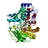
| ||||||||
|---|---|---|---|---|---|---|---|---|---|
| 1 |
| ||||||||
| Unit cell |
|
-Components
| #1: Protein | Mass: 44869.879 Da / Num. of mol.: 1 Source method: isolated from a genetically manipulated source Source: (gene. exp.) STREPTOMYCES COELICOLOR (bacteria) / Strain: A3(2) / Plasmid: PBAD/MYC-HISA / Production host: |
|---|---|
| #2: Chemical | ChemComp-FAD /   Mass: 785.550 Da / Num. of mol.: 1 / Source method: obtained synthetically / Formula: C27H33N9O15P2 / Comment: FAD*YM Mass: 785.550 Da / Num. of mol.: 1 / Source method: obtained synthetically / Formula: C27H33N9O15P2 / Comment: FAD*YM |
| #3: Chemical | ChemComp-MTL /   Mass: 182.172 Da / Num. of mol.: 1 / Source method: obtained synthetically / Formula: C6H14O6 / Comment: medication*YM Mass: 182.172 Da / Num. of mol.: 1 / Source method: obtained synthetically / Formula: C6H14O6 / Comment: medication*YM |
| #4: Water | ChemComp-HOH /  Mass: 18.015 Da / Num. of mol.: 439 / Source method: isolated from a natural source / Formula: H2O Mass: 18.015 Da / Num. of mol.: 439 / Source method: isolated from a natural source / Formula: H2O |
| Has protein modification | Y |
-Experimental details
-Experiment
| Experiment | Method: X-RAY DIFFRACTION / Number of used crystals: 1 |
|---|
- Sample preparation
| Crystal | Density Matthews: 2.05 Å3/Da / Density % sol: 39.48 % / Description: NONE |
|---|---|
| Crystal grow | pH: 6.5 / Details: pH 6.5 |
-Data collection
| Diffraction | Mean temperature: 100 K |
|---|---|
| Diffraction source | Source: ROTATING ANODE / Type: RIGAKU RU200 / Wavelength: 1.5418 |
| Detector | Date: Mar 30, 2007 |
| Radiation | Protocol: SINGLE WAVELENGTH / Monochromatic (M) / Laue (L): M / Scattering type: x-ray |
| Radiation wavelength | Wavelength: 1.5418 Å / Relative weight: 1 |
| Reflection | Resolution: 1.9→19.9 Å / Num. obs: 32193 / % possible obs: 99.9 % / Observed criterion σ(I): 0 / Redundancy: 3 % / Rmerge(I) obs: 0.1 / Net I/σ(I): 12.4 |
| Reflection shell | Resolution: 1.9→2 Å / Redundancy: 2.9 % / Rmerge(I) obs: 0.54 / Mean I/σ(I) obs: 1.9 / % possible all: 100 |
- Processing
| Software |
| ||||||||||||||||||||||||||||||||||||||||||||||||||||||||||||||||||||||||||||||||||||||||||||||||||||||||||||||||||||||||||||||||||||||||||||||||||||||||||||||||||||||||||||||||||||||
|---|---|---|---|---|---|---|---|---|---|---|---|---|---|---|---|---|---|---|---|---|---|---|---|---|---|---|---|---|---|---|---|---|---|---|---|---|---|---|---|---|---|---|---|---|---|---|---|---|---|---|---|---|---|---|---|---|---|---|---|---|---|---|---|---|---|---|---|---|---|---|---|---|---|---|---|---|---|---|---|---|---|---|---|---|---|---|---|---|---|---|---|---|---|---|---|---|---|---|---|---|---|---|---|---|---|---|---|---|---|---|---|---|---|---|---|---|---|---|---|---|---|---|---|---|---|---|---|---|---|---|---|---|---|---|---|---|---|---|---|---|---|---|---|---|---|---|---|---|---|---|---|---|---|---|---|---|---|---|---|---|---|---|---|---|---|---|---|---|---|---|---|---|---|---|---|---|---|---|---|---|---|---|---|
| Refinement | Method to determine structure: MOLECULAR REPLACEMENT Starting model: PDB ENTRY 2VFR Resolution: 1.9→19.87 Å / Cor.coef. Fo:Fc: 0.964 / Cor.coef. Fo:Fc free: 0.943 / SU B: 3.145 / SU ML: 0.092 / Cross valid method: THROUGHOUT / ESU R: 0.139 / ESU R Free: 0.132 / Stereochemistry target values: MAXIMUM LIKELIHOOD / Details: HYDROGENS HAVE BEEN ADDED IN THE RIDING POSITIONS.
| ||||||||||||||||||||||||||||||||||||||||||||||||||||||||||||||||||||||||||||||||||||||||||||||||||||||||||||||||||||||||||||||||||||||||||||||||||||||||||||||||||||||||||||||||||||||
| Solvent computation | Ion probe radii: 0.8 Å / Shrinkage radii: 0.8 Å / VDW probe radii: 1.4 Å / Solvent model: MASK | ||||||||||||||||||||||||||||||||||||||||||||||||||||||||||||||||||||||||||||||||||||||||||||||||||||||||||||||||||||||||||||||||||||||||||||||||||||||||||||||||||||||||||||||||||||||
| Displacement parameters | Biso mean: 18.11 Å2
| ||||||||||||||||||||||||||||||||||||||||||||||||||||||||||||||||||||||||||||||||||||||||||||||||||||||||||||||||||||||||||||||||||||||||||||||||||||||||||||||||||||||||||||||||||||||
| Refinement step | Cycle: LAST / Resolution: 1.9→19.87 Å
| ||||||||||||||||||||||||||||||||||||||||||||||||||||||||||||||||||||||||||||||||||||||||||||||||||||||||||||||||||||||||||||||||||||||||||||||||||||||||||||||||||||||||||||||||||||||
| Refine LS restraints |
|