 Movie
Movie Controller

[English] 日本語
 Yorodumi
- PDB-2v9a: Structure of Citrate-free Periplasmic Domain of Sensor Histidine ... -
+ Open data

- Basic information
| Entry | Database: PDB / ID: 2v9a | ||||||
|---|---|---|---|---|---|---|---|
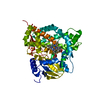
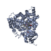
| Title | Structure of Citrate-free Periplasmic Domain of Sensor Histidine Kinase CitA | ||||||
 Components Components | SENSOR KINASE CITA | ||||||
 Keywords Keywords | TRANSFERASE / SIGNAL TRANSDUCTION / SENSOR HISTIDINE KINASE / CITA / KINASE / MEMBRANE / SENSOR DOMAIN / TRANSMEMBRANE / INNER MEMBRANE / PHOSPHORYLATION / TWO-COMPONENT REGULATORY SYSTEM | ||||||
| Function / homology |  Function and homology information Function and homology informationphosphorelay sensor kinase activity / histidine kinase / ATP binding / plasma membrane Similarity search - Function | ||||||
| Biological species |  KLEBSIELLA PNEUMONIAE (bacteria) KLEBSIELLA PNEUMONIAE (bacteria) | ||||||
| Method |  X-RAY DIFFRACTION / SYNCHROTRON / MOLECULAR REPLACEMENT / Resolution: 2 Å X-RAY DIFFRACTION / SYNCHROTRON / MOLECULAR REPLACEMENT / Resolution: 2 Å | ||||||
 Authors Authors | Sevvana, M. / Vijayan, V. / Zweckstetter, M. / Reinelt, S. / Madden, D.R. / Sheldrick, G.M. / Bott, M. / Griesinger, C. / Becker, S. | ||||||
 Citation Citation | Journal: J.Mol.Biol. / Year: 2008 Title: A Ligand-Induced Switch in the Periplasmic Domain of Sensor Histidine Kinase Cita. Authors: Sevvana, M. / Vijayan, V. / Zweckstetter, M. / Reinelt, S. / Madden, D.R. / Herbst-Irmer, R. / Sheldrick, G.M. / Bott, M. / Griesinger, C. / Becker, S. | ||||||
| History |
|
- Structure visualization
| Structure viewer | Molecule: MolmilJmol/JSmol |
|---|
- Downloads & links
-Download
| PDBx/mmCIF format | 2v9a.cif.gz | 56.3 KB | Display | PDBx/mmCIF format |
|---|---|---|---|---|
| PDB format | pdb2v9a.ent.gz | 40.2 KB | Display | PDB format |
| PDBx/mmJSON format | 2v9a.json.gz | Tree view | PDBx/mmJSON format | |
| Others |  Other downloads Other downloads |
-Validation report
| Arichive directory | https://data.pdbj.org/pub/pdb/validation_reports/v9/2v9aftp://data.pdbj.org/pub/pdb/validation_reports/v9/2v9a | HTTPS FTP |
|---|
-Related structure data
| Related structure data |  2j80SC S: Starting model for refinement C: citing same article ( |
|---|---|
| Similar structure data |


-Links
 PDBj
PDBj

- Assembly
| Deposited unit | 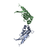
| ||||||||
|---|---|---|---|---|---|---|---|---|---|
| 1 | 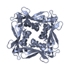
| ||||||||
| 2 | 
| ||||||||
| Unit cell |
|
-Components
| #1: Protein | Mass: 14499.438 Da / Num. of mol.: 2 Fragment: PERIPLASMIC LIGAND BINDING DOMAIN, RESIDUES 45-176 Source method: isolated from a genetically manipulated source Source: (gene. exp.) KLEBSIELLA PNEUMONIAE (bacteria) / Plasmid: PET16B / Production host: #2: Water | ChemComp-HOH / |  Mass: 18.015 Da / Num. of mol.: 229 / Source method: isolated from a natural source / Formula: H2O Mass: 18.015 Da / Num. of mol.: 229 / Source method: isolated from a natural source / Formula: H2O |
|---|
-Experimental details
-Experiment
| Experiment | Method: X-RAY DIFFRACTION / Number of used crystals: 1 |
|---|
- Sample preparation
| Crystal | Density Matthews: 2.38 Å3/Da / Density % sol: 48.3 % / Description: MEROHEDRAL TWINNING ALONG 110 |
|---|---|
| Crystal grow | Temperature: 293 K / Method: vapor diffusion, hanging drop / pH: 7.5 Details: HANGING DROP, VAPOUR DIFFUSION, RESERVOIR: 20MM HEPES,PH 7.5,0.63M NAH2PO4,0.63M KH2PO4, PROTEIN SOLUTION: 15 MG/ML,MIXING RATIO 1:1,TEMPERATURE 293K |
-Data collection
| Diffraction | Mean temperature: 100 K |
|---|---|
| Diffraction source | Source: SYNCHROTRON / Site: MPG/DESY, HAMBURG  / Beamline: BW6 / Wavelength: 1 / Beamline: BW6 / Wavelength: 1 |
| Detector | Type: MARRESEARCH / Detector: CCD / Date: Mar 8, 2006 / Details: MIRROR |
| Radiation | Protocol: SINGLE WAVELENGTH / Monochromatic (M) / Laue (L): M / Scattering type: x-ray |
| Radiation wavelength | Wavelength: 1 Å / Relative weight: 1 |
| Reflection | Resolution: 2→25 Å / Num. obs: 19151 / % possible obs: 97 % / Observed criterion σ(I): 2 / Redundancy: 5.49 % / Rmerge(I) obs: 0.05 / Net I/σ(I): 16.7 |
| Reflection shell | Resolution: 2→2.1 Å / Redundancy: 1.6 % / Rmerge(I) obs: 0.19 / % possible all: 84.1 |
- Processing
| Software |
| ||||||||||||||||
|---|---|---|---|---|---|---|---|---|---|---|---|---|---|---|---|---|---|
| Refinement | Method to determine structure: MOLECULAR REPLACEMENT Starting model: PDB ENTRY 2J80 Resolution: 2→24.6626 Å Details: THE STRUCTURE WAS FIRST REFINED WITH CNS, FOLLOWED BY SHELXL AND FOR BETTER CONVERGENCE WITH PHENIX.REFINE TWIN REFINEMENT PROTOCOL TWIN LAW 1 0 0 0 -1 0 0 0 -1 TWIN FRACTION 0.40
| ||||||||||||||||
| Refinement step | Cycle: LAST / Resolution: 2→24.6626 Å
|
