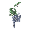
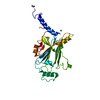
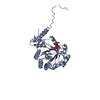
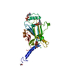
- PDB-2uv3: Structure of the signal-regulatory protein (SIRP) alpha domain th... -
+
Open data
ID or keywords:
Loading...
-
Basic information
Entry
Database: PDB / ID: 2uv3
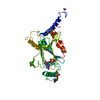
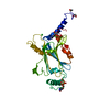
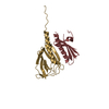
Title
Structure of the signal-regulatory protein (SIRP) alpha domain that binds CD47.
Components
TYROSINE-PROTEIN PHOSPHATASE NON-RECEPTOR TYPE SUBSTRATE 1
Keywords
RECEPTOR / CD47-BINDING DOMAIN OF SIRP-ALPHA / MEMBRANE / SH3- BINDING / GLYCOPROTEIN / TRANSMEMBRANE / PHOSPHORYLATION / HUMAN SIRP-ALPHA N TERMINAL V DOMAIN / IMMUNOGLOBULIN DOMAIN / SIGNAL-REGULATORY PROTEIN ALPHA
Function / homology
Function and homology information
cellular response to interleukin-12 / monocyte extravasation / negative regulation of macrophage inflammatory protein 1 alpha production / protein binding involved in heterotypic cell-cell adhesion / negative regulation of chemokine (C-C motif) ligand 5 production / regulation of interleukin-1 beta production / GTPase regulator activity / cell-cell adhesion mediator activity / regulation of type II interferon production / negative regulation of lipopolysaccharide-mediated signaling pathway ...cellular response to interleukin-12 / monocyte extravasation / negative regulation of macrophage inflammatory protein 1 alpha production / protein binding involved in heterotypic cell-cell adhesion / negative regulation of chemokine (C-C motif) ligand 5 production / regulation of interleukin-1 beta production / GTPase regulator activity / cell-cell adhesion mediator activity / regulation of type II interferon production / negative regulation of lipopolysaccharide-mediated signaling pathway / negative regulation of nitric oxide biosynthetic process / regulation of tumor necrosis factor production / protein antigen binding / negative regulation of interferon-beta production / regulation of nitric oxide biosynthetic process / regulation of interleukin-6 production / Signal regulatory protein family interactions / negative regulation of JNK cascade / protein phosphatase inhibitor activity / negative regulation of phagocytosis / negative regulation of interleukin-6 production / tertiary granule membrane / ficolin-1-rich granule membrane / negative regulation of tumor necrosis factor production / cellular response to interleukin-1 / negative regulation of cytokine production involved in inflammatory response / protein tyrosine kinase binding / positive regulation of phagocytosis / negative regulation of canonical NF-kappaB signal transduction / Cell surface interactions at the vascular wall / negative regulation of ERK1 and ERK2 cascade / SH3 domain binding / negative regulation of inflammatory response / cellular response to type II interferon / positive regulation of reactive oxygen species metabolic process / cellular response to hydrogen peroxide / positive regulation of T cell activation / cell migration / regulation of gene expression / protein phosphatase binding / cell adhesion / Neutrophil degranulation / cell surface / extracellular exosome / membrane / plasma membrane Similarity search - Function
SHEET THE SHEET STRUCTURE OF THIS MOLECULE IS BIFURCATED. IN ORDER TO REPRESENT THIS FEATURE IN ... SHEET THE SHEET STRUCTURE OF THIS MOLECULE IS BIFURCATED. IN ORDER TO REPRESENT THIS FEATURE IN THE SHEET RECORDS BELOW, TWO SHEETS ARE DEFINED.
In the structure databanks used in Yorodumi, some data are registered as the other names, "COVID-19 virus" and "2019-nCoV". Here are the details of the virus and the list of structure data.
Jan 31, 2019. EMDB accession codes are about to change! (news from PDBe EMDB page)
EMDB accession codes are about to change! (news from PDBe EMDB page)
The allocation of 4 digits for EMDB accession codes will soon come to an end. Whilst these codes will remain in use, new EMDB accession codes will include an additional digit and will expand incrementally as the available range of codes is exhausted. The current 4-digit format prefixed with “EMD-” (i.e. EMD-XXXX) will advance to a 5-digit format (i.e. EMD-XXXXX), and so on. It is currently estimated that the 4-digit codes will be depleted around Spring 2019, at which point the 5-digit format will come into force.
The EM Navigator/Yorodumi systems omit the EMD- prefix.
Related info.:Q: What is EMD? / ID/Accession-code notation in Yorodumi/EM Navigator
Yorodumi is a browser for structure data from EMDB, PDB, SASBDB, etc.
This page is also the successor to EM Navigator detail page, and also detail information page/front-end page for Omokage search.
The word "yorodu" (or yorozu) is an old Japanese word meaning "ten thousand". "mi" (miru) is to see.
Related info.:EMDB / PDB / SASBDB / Comparison of 3 databanks / Yorodumi Search / Aug 31, 2016. New EM Navigator & Yorodumi / Yorodumi Papers / Jmol/JSmol / Function and homology information / Changes in new EM Navigator and Yorodumi
 Movie
Movie Controller

 Yorodumi
 Open data
 Basic information
 Components
 Keywords
 Function and homology information
 HOMO SAPIENS (human)
 X-RAY DIFFRACTION /
  Authors
 Citation
 Structure visualization
 Downloads & links
 Other downloads
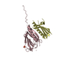
 PDBj
PDBj

 Assembly
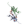

 CRICETULUS GRISEUS (Chinese hamster) / Variant (production host): LEC3.2.8.1 / References: UniProt: P78324
 Mass: 195.237 Da / Num. of mol.: 2 / Source method: obtained synthetically / Formula: C6H13NO4S / Comment: pH buffer*YM
 Mass: 96.063 Da / Num. of mol.: 6 / Source method: obtained synthetically / Formula: SO4
 Mass: 18.015 Da / Num. of mol.: 298 / Source method: isolated from a natural source / Formula: H2O
 Sample preparation
 / Beamline: BM14 / Wavelength: 0.8856, 0.9783, 0.9079, 0.9792
 Processing