 Movie
Movie Controller


+ Open data

- Basic information
| Entry | Database: PDB / ID: 2rta | |||||||||
|---|---|---|---|---|---|---|---|---|---|---|
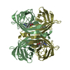
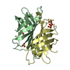
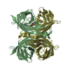
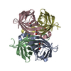
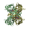
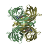
| Title | APOSTREPTAVIDIN, PH 2.97, SPACE GROUP I4122 | |||||||||
 Components Components | STREPTAVIDIN | |||||||||
 Keywords Keywords | BIOTIN-BINDING PROTEIN / I4122 APOSTREPTAVIDIN / PH 2.97 | |||||||||
| Function / homology |  Function and homology information Function and homology information | |||||||||
| Biological species |  Streptomyces avidinii (bacteria) Streptomyces avidinii (bacteria) | |||||||||
| Method | X-RAY DIFFRACTION / Resolution: 1.39 Å | |||||||||
 Authors Authors | Katz, B.A. | |||||||||
 Citation Citation | Journal: J.Mol.Biol. / Year: 1997 Title: Binding of biotin to streptavidin stabilizes intersubunit salt bridges between Asp61 and His87 at low pH. Authors: Katz, B.A. #1: Journal: J.Biol.Chem. / Year: 1997Title: In Crystals of Complexes of Streptavidin with Peptide Ligands Containing the Hpq Sequence the Pka of the Peptide Histidine is Less Than 3.0 Authors: Katz, B.A. / Cass, R.T. #2: Journal: J.Am.Chem.Soc. / Year: 1996Title: Structure-Based Design Tools: Structural and Thermodynamic Comparison with Biotin of a Small Molecule that Binds Streptavidin with Micromolar Affinity Authors: Katz, B.A. / Liu, B. / Cass, R.T. #3: Journal: J.Am.Chem.Soc. / Year: 1996Title: Preparation of a Protein-Dimerizing Ligand by Topochemistry and Structure-Based Design Authors: Katz, B.A. #4: Journal: J.Biol.Chem. / Year: 1995Title: Topochemical Catalysis Achieved by Structure-Based Ligand Design Authors: Katz, B.A. / Cass, R.T. / Liu, B. / Arze, R. / Collins, N. #5: Journal: Chem.Biol. / Year: 1995Title: Topochemistry for Preparing Ligands that Dimerize Receptors Authors: Katz, B.A. / Stroud, R.M. / Collins, N. / Liu, B. / Arze, R. #6: Journal: Biochemistry / Year: 1995Title: Binding to Protein Targets of Peptidic Leads Discovered by Phage Display: Crystal Structures of Streptavidin-Bound Linear and Cyclic Peptide Ligands Containing the Hpq Sequence Authors: Katz, B.A. #7: Journal: J.Am.Chem.Soc. / Year: 1995Title: Structure-Based Design of High Affinity Streptavidin Binding Cyclic Peptide Ligands Containing Thioether Cross-Links Authors: Katz, B.A. / Johnson, C.R. / Cass, R.T. | |||||||||
| History |
|
- Structure visualization
| Structure viewer | Molecule: MolmilJmol/JSmol |
|---|
- Downloads & links
-Download
| PDBx/mmCIF format | 2rta.cif.gz | 63.2 KB | Display | PDBx/mmCIF format |
|---|---|---|---|---|
| PDB format | pdb2rta.ent.gz | 49.4 KB | Display | PDB format |
| PDBx/mmJSON format | 2rta.json.gz | Tree view | PDBx/mmJSON format | |
| Others |  Other downloads Other downloads |
-Validation report
| Arichive directory | https://data.pdbj.org/pub/pdb/validation_reports/rt/2rtaftp://data.pdbj.org/pub/pdb/validation_reports/rt/2rta | HTTPS FTP |
|---|
-Related structure data
| Related structure data |  2izaC  2izbC  2izcC 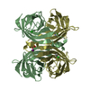 2izdC  2izeC  2izfC  2izgC  2izhC  2iziC  2izjC  2izkC 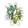 2izlC  2rtbC  2rtcC  2rtdC  2rteC  2rtfC  2rtgC  2rthC  2rtiC 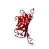 2rtjC  2rtkC  2rtlC  2rtmC  2rtnC  2rtoC 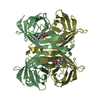 2rtpC  2rtqC  2rtrC C: citing same article ( |
|---|---|
| Similar structure data |


-Links
 PDBj
PDBj- Assembly
| Deposited unit | 
| ||||||||||||
|---|---|---|---|---|---|---|---|---|---|---|---|---|---|
| 1 | 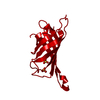
| ||||||||||||
| Unit cell |
| ||||||||||||
| Components on special symmetry positions |
|
-Components
| #1: Protein | Mass: 14181.324 Da / Num. of mol.: 1 / Source method: isolated from a natural source / Source: (natural) Streptomyces avidinii (bacteria) / References: UniProt: P22629 | ||
|---|---|---|---|
| #2: Chemical | Sulfate  Mass: 96.063 Da / Num. of mol.: 2 / Source method: obtained synthetically / Formula: SO4 Mass: 96.063 Da / Num. of mol.: 2 / Source method: obtained synthetically / Formula: SO4#3: Water | ChemComp-HOH / | Water Mass: 18.015 Da / Num. of mol.: 53 / Source method: isolated from a natural source / Formula: H2O Mass: 18.015 Da / Num. of mol.: 53 / Source method: isolated from a natural source / Formula: H2O |
-Experimental details
-Experiment
| Experiment | Method: X-RAY DIFFRACTION / Number of used crystals: 1 |
|---|
- Sample preparation
| Crystal | Density Matthews: 2.89 Å3/Da / Density % sol: 37.1 % Description: REJECTION CRITERIA: (I(H)I - ) > [0.30 * () + 0.10*I(H)I], WHERE I(H)I IS THE ITH OBSERVATION OF THE INTENSITY OF REFLECTION H (M.G.ROSSMANN, A.G.W.LESLIE, S.S.ABDEL-MEGUID, T.TSUKIHARA, ...Description: REJECTION CRITERIA: (I(H)I - | ||||||||||||||||||||||||||||||
|---|---|---|---|---|---|---|---|---|---|---|---|---|---|---|---|---|---|---|---|---|---|---|---|---|---|---|---|---|---|---|---|
| Crystal grow | pH: 2.97 Details: SYNTHETIC MOTHER LIQUOR OF 75% SATURATED AMMONIUM SULFATE, 25% 1.0 M SODIUM FORMATE ADJUSTED TO PH 2.97. | ||||||||||||||||||||||||||||||
| Crystal | *PLUS | ||||||||||||||||||||||||||||||
| Crystal grow | *PLUS Temperature: 20 ℃ / pH: 4.5 / Method: vapor diffusion, hanging drop / Details: Pahler, A., (1987) J. Biol. Chem., 262, 13933. | ||||||||||||||||||||||||||||||
| Components of the solutions | *PLUS
|
-Data collection
| Diffraction | Mean temperature: 273 K |
|---|---|
| Diffraction source | Wavelength: 1.5418 |
| Detector | Type: RIGAKU RAXIS IV / Detector: IMAGE PLATE |
| Radiation | Monochromatic (M) / Laue (L): M / Scattering type: x-ray |
| Radiation wavelength | Wavelength: 1.5418 Å / Relative weight: 1 |
| Reflection | Num. obs: 30482 / Redundancy: 4.1 % / Rmerge(I) obs: 0.066 |
| Reflection | *PLUS Highest resolution: 1.32 Å / Num. measured all: 123910 |
- Processing
| Software |
| ||||||||||||||||||||||||||||||||||||||||||||||||||||||||||||
|---|---|---|---|---|---|---|---|---|---|---|---|---|---|---|---|---|---|---|---|---|---|---|---|---|---|---|---|---|---|---|---|---|---|---|---|---|---|---|---|---|---|---|---|---|---|---|---|---|---|---|---|---|---|---|---|---|---|---|---|---|---|
| Refinement | Resolution: 1.39→7.5 Å / σ(F): 1.8 Details: THE FOLLOWING ATOMS HAD WEAK DENSITY AND OCCUPANCIES WERE REFINED: ALA 13, GLU 14, ALA 15 (EXCEPT FOR C AND O), GLN 24, LEU 25, GLY 26 ALA 35 (SIDE CHAIN), ASP 36 (N, HN, CA, HA, C, O, CB, ...Details: THE FOLLOWING ATOMS HAD WEAK DENSITY AND OCCUPANCIES WERE REFINED: ALA 13, GLU 14, ALA 15 (EXCEPT FOR C AND O), GLN 24, LEU 25, GLY 26 ALA 35 (SIDE CHAIN), ASP 36 (N, HN, CA, HA, C, O, CB, HB1, HB2, CG, OD1,OD2), GLU 44 (CG, HG1, HG2, CD, OE1, OE2) SER 45 (MAIN CHAIN), ALA 46 (CA, HA, C, O, CB, HB1, HB2, HB3), VAL 47, GLY 48 ASN 49, ALA 50, GLU 51, SER 52 (SIDE CHAIN), ARG 53 (NE, HE, CZ, NH1, HH11, HH12, NH2, HH21, HH22) ARG 84 (CG, HG1, HG2, CD, HD1, HD2, NE, HE, CZ, NH1, HH11, HH12, NH2, HH21, HH22) GLY 99, ALA 100 (MAIN CHAIN), ALA 100 (SIDE CHAIN), GLU 101 (N, HN, CA, HA, CB, HB1, HB2, C, O, CG, HG1, HG2, CD, OE1, OE2) ARG 103 (NE, HE, CZ, NH1, HH11, HH12, NH2, HH21, HH22), GLU 116 (N, HN, CA, HA, CB, HB1, HB2, CG, HG1, HG2, C, O, CD, OE1, OE2) ALA 117, ASN 118 (SIDE CHAIN), LYS 121 (CG, HG1, HG2, CD, HD1, HD2, CE, HE1, HE2, NZ, HZ1,HZ2, HZ3) RESIDUES TYR 60 TO SER 69 WERE REFINED IN 2 CONFORMATIONS BECAUSE UPON PROTONATION OF ASP 61 AT LOW PH, ASP 61 UNDERGOES A LARGE SHIFT IN CONFORMATION AND CHANGE IN HYDROGEN BONDING. THE LOOP COMPRISING RESIDUES ASP 61 TO SER 69 ALSO UNDERGO CORRESPONDING CONFORMATIONAL CHANGES. HOWEVER SOME OF THESE RESIDUES ARE DISORDERED AND NOT VISIBLE IN EITHER CONFORMATION. DISCRETELY DISORDERED SIDE CHAINS WHOSE OCCUPANCIES AND STRUCTURES WERE SIMULTANEOUSLY REFINED ARE: SER 45, LEU 73, HIS 87, GLN 107, LEU 110, LYS 132. DISORDERED SOLVENTS ARE: HOH 134 WHICH OCCUPIES THE SPACE AVAILABLE WHEN ASP 61 IS IN CONFORMATION NUMBER 2 HOH 142 WHICH IS CLOSE TO A SYMMETRY-RELATED EQUIVALENT OF ITSELF HOH 150 WHICH IS CLOSE TO HOH 151 SO4 153 WHICH IS CLOSE TO HOH 545 HOH 152 WHICH IS CLOSE TO SO4 154 HOH 207 WHICH IS CLOSE TO A SYMMETRY-RELATED EQUIVALENT OF HOH 354 HOH 305 WHICH IS CLOSE TO A SYMMETRY-RELATED EQUIVALENT OF ITSELF HOH 546 WHICH IS CLOSE TO A SYMMETRY-RELATED EQUIVALENT OF ITSELF HOH 550 WHICH IS CLOSE TO A SYMMETRY-RELATED EQUIVALENT OF ITSELF HOH 635 WHICH IS CLOSE TO A SYMMETRY-RELATED EQUIVALENT OF ITSELF THE FOLLOWING ATOMS HAD WEAK DENSITY AND OCCUPANCIES WERE REFINED: ALA 13 GLU 14 ALA 15 (EXCEPT FOR C AND O) GLN 24 LEU 25 GLY 26 ALA 35 (SIDE CHAIN) ASP 36 (N, HN, CA, HA, C, O, CB, HB1, HB2, CG, OD1,OD2) GLU 44 (CG, HG1, HG2, CD, OE1, OE2) SER 45 (MAIN CHAIN) ALA 46 (CA, HA, C, O, CB, HB1, HB2, HB3) VAL 47 GLY 48 ASN 49 ALA 50 GLU 51 SER 52 (SIDE CHAIN) ARG 53 (NE, HE, CZ, NH1, HH11, HH12, NH2, HH21, HH22) ARG 84 (CG, HG1, HG2, CD, HD1, HD2, NE, HE, CZ, NH1, HH11, HH12, NH2, HH21, HH22) GLY 99 ALA 100 (MAIN CHAIN) ALA 100 (SIDE CHAIN) GLU 101 (N, HN, CA, HA, CB, HB1, HB2, C, O, CG, HG1, HG2, CD, OE1, OE2) ARG 103 (NE, HE, CZ, NH1, HH11, HH12, NH2, HH21, HH22) GLU 116 (N, HN, CA, HA, CB, HB1, HB2, CG, HG1, HG2, C, O, CD, OE1, OE2) ALA 117 ASN 118 (SIDE CHAIN) LYS 121 (CG, HG1, HG2, CD, HD1, HD2, CE, HE1, HE2, NZ, HZ1, HZ2, HZ3) RESIDUES TYR 60 TO SER 69 WERE REFINED IN 2 CONFORMATIONS BECAUSE UPON PROTONATION OF ASP 61 AT LOW PH, ASP 61 UNDERGOES A LARGE SHIFT IN CONFORMATION AND CHANGE IN HYDROGEN BONDING. THE LOOP COMPRISING RESIDUES ASP 61 TO SER 69 ALSO UNDERGO CORRESPONDING CONFORMATIONAL CHANGES. HOWEVER SOME OF THESE RESIDUES ARE DISORDERED AND NOT VISIBLE IN EITHER CONFORMATION.
| ||||||||||||||||||||||||||||||||||||||||||||||||||||||||||||
| Refinement step | Cycle: LAST / Resolution: 1.39→7.5 Å
| ||||||||||||||||||||||||||||||||||||||||||||||||||||||||||||
| Refine LS restraints |
| ||||||||||||||||||||||||||||||||||||||||||||||||||||||||||||
| LS refinement shell | Resolution: 1.39→1.45 Å / % reflection obs: 30.1 % | ||||||||||||||||||||||||||||||||||||||||||||||||||||||||||||
| Software | *PLUS Name: X-PLOR / Classification: refinement | ||||||||||||||||||||||||||||||||||||||||||||||||||||||||||||
| Refine LS restraints | *PLUS
| ||||||||||||||||||||||||||||||||||||||||||||||||||||||||||||
| LS refinement shell | *PLUS Rfactor obs: 0.206 |
