 Movie
Movie Controller

[English] 日本語
 Yorodumi
- PDB-2qtx: Crystal structure of an Hfq-like protein from Methanococcus jannaschii -
+ Open data

- Basic information
| Entry | Database: PDB / ID: 2qtx | ||||||
|---|---|---|---|---|---|---|---|
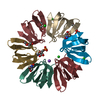
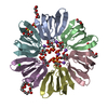
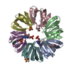
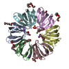
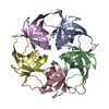
| Title | Crystal structure of an Hfq-like protein from Methanococcus jannaschii | ||||||
 Components Components | Uncharacterized protein MJ1435 | ||||||
 Keywords Keywords | RNA BINDING PROTEIN / Hfq / Sm / RNA-binding protein / sRNA / translational regulation | ||||||
| Function / homology |  Function and homology information Function and homology information | ||||||
| Biological species |   Methanocaldococcus jannaschii (archaea) Methanocaldococcus jannaschii (archaea) | ||||||
| Method |  X-RAY DIFFRACTION / SYNCHROTRON / MOLECULAR REPLACEMENT / molecular replacement / Resolution: 2.5 Å X-RAY DIFFRACTION / SYNCHROTRON / MOLECULAR REPLACEMENT / molecular replacement / Resolution: 2.5 Å | ||||||
 Authors Authors | Nielsen, J.S. / Boggild, A. / Andersen, C.B.F. / Nielsen, G. / Boysen, A. / Brodersen, D.E. / Valentin-Hansen, P. | ||||||
 Citation Citation | Journal: Rna / Year: 2007 Title: An Hfq-like protein in archaea: Crystal structure and functional characterization of the Sm protein from Methanococcus jannaschii. Authors: Nielsen, J.S. / Boggild, A. / Andersen, C.B. / Nielsen, G. / Boysen, A. / Brodersen, D.E. / Valentin-Hansen, P. | ||||||
| History |
|
- Structure visualization
| Structure viewer | Molecule: MolmilJmol/JSmol |
|---|
- Downloads & links
-Download
| PDBx/mmCIF format | 2qtx.cif.gz | 287 KB | Display | PDBx/mmCIF format |
|---|---|---|---|---|
| PDB format | pdb2qtx.ent.gz | 235 KB | Display | PDB format |
| PDBx/mmJSON format | 2qtx.json.gz | Tree view | PDBx/mmJSON format | |
| Others |  Other downloads Other downloads |
-Validation report
| Arichive directory | https://data.pdbj.org/pub/pdb/validation_reports/qt/2qtxftp://data.pdbj.org/pub/pdb/validation_reports/qt/2qtx | HTTPS FTP |
|---|
-Related structure data
| Related structure data | 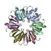 1u1sS S: Starting model for refinement |
|---|---|
| Similar structure data |


-Links
 PDBj
PDBj

- Assembly
| Deposited unit | 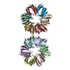
| ||||||||
|---|---|---|---|---|---|---|---|---|---|
| 1 | 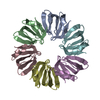
| ||||||||
| 2 | 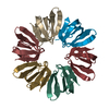
| ||||||||
| Unit cell |
| ||||||||
| Details | The biological assembly is a homohexamer. The asu contains two hexamers. |
-Components
| #1: Protein | Mass: 8309.771 Da / Num. of mol.: 12 Source method: isolated from a genetically manipulated source Source: (gene. exp.) Methanocaldococcus jannaschii (archaea)Plasmid: pTYP11 / Production host:  #2: Water | ChemComp-HOH / |  Mass: 18.015 Da / Num. of mol.: 130 / Source method: isolated from a natural source / Formula: H2O Mass: 18.015 Da / Num. of mol.: 130 / Source method: isolated from a natural source / Formula: H2O |
|---|
-Experimental details
-Experiment
| Experiment | Method: X-RAY DIFFRACTION / Number of used crystals: 1 |
|---|
- Sample preparation
| Crystal | Density Matthews: 2.25 Å3/Da / Density % sol: 45.31 % |
|---|---|
| Crystal grow | Temperature: 277 K / pH: 8.5 Details: 25% PEG 3350, 0.2M Ammonium Acetate, 0.1M Tris, VAPOR DIFFUSION, SITTING DROP, temperature 277K, pH 8.50 |
-Data collection
| Diffraction | Mean temperature: 100 K | |||||||||||||||||||||||||||||||||||||||||||||||||||||||||||||||||||||||||||||
|---|---|---|---|---|---|---|---|---|---|---|---|---|---|---|---|---|---|---|---|---|---|---|---|---|---|---|---|---|---|---|---|---|---|---|---|---|---|---|---|---|---|---|---|---|---|---|---|---|---|---|---|---|---|---|---|---|---|---|---|---|---|---|---|---|---|---|---|---|---|---|---|---|---|---|---|---|---|---|
| Diffraction source | Source: SYNCHROTRON / Site: SLS  / Beamline: X06SA / Wavelength: 0.9993 / Beamline: X06SA / Wavelength: 0.9993 | |||||||||||||||||||||||||||||||||||||||||||||||||||||||||||||||||||||||||||||
| Detector | Type: MARMOSAIC 225 mm CCD / Detector: CCD / Date: Nov 17, 2006 / Details: DYNAMICALLY BENDABLE MIRRORS | |||||||||||||||||||||||||||||||||||||||||||||||||||||||||||||||||||||||||||||
| Radiation | Monochromator: FIXED EXIT SI 111 MONCHROMATOR / Protocol: SINGLE WAVELENGTH / Monochromatic (M) / Laue (L): M / Scattering type: x-ray | |||||||||||||||||||||||||||||||||||||||||||||||||||||||||||||||||||||||||||||
| Radiation wavelength | Wavelength: 0.9993 Å / Relative weight: 1 | |||||||||||||||||||||||||||||||||||||||||||||||||||||||||||||||||||||||||||||
| Reflection | Redundancy: 3.4 % / Av σ(I) over netI: 17 / Number: 96326 / Rmerge(I) obs: 0.056 / Χ2: 1.5 / D res high: 2.5 Å / D res low: 250 Å / Num. obs: 28424 / % possible obs: 92.5 | |||||||||||||||||||||||||||||||||||||||||||||||||||||||||||||||||||||||||||||
| Diffraction reflection shell |
| |||||||||||||||||||||||||||||||||||||||||||||||||||||||||||||||||||||||||||||
| Reflection | Resolution: 2.5→250 Å / Num. obs: 28424 / % possible obs: 92.5 % / Redundancy: 3.4 % / Biso Wilson estimate: 48.18 Å2 / Rmerge(I) obs: 0.056 / Net I/σ(I): 17 | |||||||||||||||||||||||||||||||||||||||||||||||||||||||||||||||||||||||||||||
| Reflection shell | Resolution: 2.5→2.59 Å / Redundancy: 1.8 % / Rmerge(I) obs: 0.209 / Mean I/σ(I) obs: 3.7 / % possible all: 57.5 |
-Phasing
| Phasing | Method: molecular replacement | |||||||||||||||||||||||||||||||||||||||||||||||||
|---|---|---|---|---|---|---|---|---|---|---|---|---|---|---|---|---|---|---|---|---|---|---|---|---|---|---|---|---|---|---|---|---|---|---|---|---|---|---|---|---|---|---|---|---|---|---|---|---|---|---|
| Phasing MR |
| |||||||||||||||||||||||||||||||||||||||||||||||||
| Phasing dm | FOM : 0.62 / FOM acentric: 0.62 / FOM centric: 0.62 / Reflection: 28398 / Reflection acentric: 27025 / Reflection centric: 1373 | |||||||||||||||||||||||||||||||||||||||||||||||||
| Phasing dm shell |
|
- Processing
| Software |
| ||||||||||||||||||||||||||||||||||||||||||||||||||||||||||||
|---|---|---|---|---|---|---|---|---|---|---|---|---|---|---|---|---|---|---|---|---|---|---|---|---|---|---|---|---|---|---|---|---|---|---|---|---|---|---|---|---|---|---|---|---|---|---|---|---|---|---|---|---|---|---|---|---|---|---|---|---|---|
| Refinement | Method to determine structure: MOLECULAR REPLACEMENT Starting model: PDB ENTRY 1U1S Resolution: 2.5→38.5 Å / Isotropic thermal model: ISOTROPIC / Cross valid method: THROUGHOUT / Stereochemistry target values: Engh & Huber
| ||||||||||||||||||||||||||||||||||||||||||||||||||||||||||||
| Solvent computation | Bsol: 30.003 Å2 / ksol: 0.326 e/Å3 | ||||||||||||||||||||||||||||||||||||||||||||||||||||||||||||
| Displacement parameters | Biso mean: 41.43 Å2
| ||||||||||||||||||||||||||||||||||||||||||||||||||||||||||||
| Refinement step | Cycle: LAST / Resolution: 2.5→38.5 Å
| ||||||||||||||||||||||||||||||||||||||||||||||||||||||||||||
| Refine LS restraints |
| ||||||||||||||||||||||||||||||||||||||||||||||||||||||||||||
| LS refinement shell | Resolution: 2.5→2.59 Å / Total num. of bins used: 54
|
